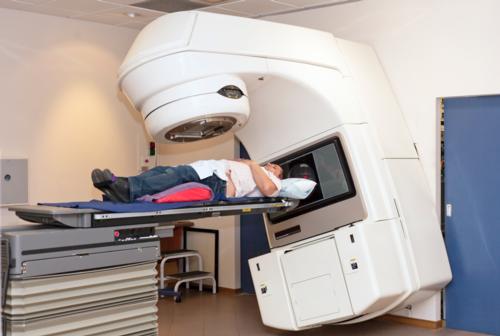
External Beam Therapy (EBT)
External beam therapy (EBT) is a method for delivering high-energy x-ray or electron beams to a patient's tumor. Beams are usually generated by a linear accelerator and targeted to destroy cancer cells while sparing surrounding normal tissues. EBT also may be used to relieve symptoms in patients with advanced cancer or cancer that has metastasized.
To prepare for EBT, your doctor will perform a physical exam and use CT scanning to conduct a treatment simulation session. Other imaging procedures may be used to help determine the exact shape and location of your tumor, and a special device may be created to help you maintain the same exact position during each treatment. Your doctor will give you specific instructions based on the treatment technique to be used.
- What is external beam therapy and how is it used?
- Who will be involved in this procedure?
- What equipment is used?
- Who operates the equipment?
- Is there any special preparation needed for the procedure?
- How is the procedure performed?
- What will I feel during this procedure?
- What kind of treatment follow-up should I expect?
What is external beam therapy and how is it used?
External beam therapy (EBT), also called external radiation therapy, is a method for delivering a beam or several beams of high-energy x-rays to a patient's tumor. Beams are generated outside the patient (usually by a linear accelerator, see below) and are targeted at the tumor site. These high energy x-rays can deposit their dose to the area of the tumor to destroy the cancer cells and, with careful treatment planning, spare the surrounding normal tissues. No radioactive sources are placed inside the patient's body.
External beam therapy is used to treat the following diseases as well as many others:
- Breast Cancer Treatment
- Colorectal (Bowel) Cancer Treatment
- Esophageal Cancer Treatment
- Head and Neck Cancer Treatment
- Lung Cancer Treatment
- Prostate Cancer Treatment
- Brain Tumor Treatment
Why is this procedure performed?
External beam therapy is most commonly used to treat cancer. Often, the goal is to eliminate a tumor or prevent a tumor from returning. The procedure may also be performed before or after surgery to remove a cancerous tumor, to reduce the tumor size before surgery, or to prevent the tumor from coming back after surgery.
EBT may also be used as a palliative treatment in patients with advanced stage cancer or cancer that has metastasized. In this case, the goal of therapy is to reduce a patient's symptoms rather than cure the cancer.
Who will be involved in this procedure?
Delivery of external beam therapy requires a treatment team, including a radiation oncologist, medical physicist, dosimetrist and radiation therapist. The radiation oncologist is a physician who evaluates the patient and determines the appropriate therapy or combination of therapies. He or she determines what area to treat and what dose to deliver. Together with the medical physicist and the dosimetrist, the radiation oncologist determines what techniques to use to deliver the prescribed dose. The physicist and the dosimetrist then make detailed treatment calculations and quality assurance checks prior to treatment delivery. The radiation therapists are specially trained technologists who deliver the daily treatments.
What equipment is used?
Radiation oncologists use linear accelerators or cobalt machines to deliver external beam therapy. Your radiation oncologist will determine the equipment most suited to your treatment. The linear accelerator is the most commonly used device for external beam therapy.
- Linear Accelerator - see linear accelerator page
Who operates the equipment?
The equipment is operated by a radiation therapist, a highly trained technologist. The overall treatment plan is created by the radiation oncologist, a highly trained physician specializing in treating cancer with radiotherapy.
Is there any special preparation needed for the procedure?
The process of external beam therapy involves three parts:
- Simulation
- Treatment Planning
- Treatment Delivery
The goal of simulation is to determine the treatment position that will be used daily, to make devices that will help the patient maintain that position, and to obtain the necessary images for treatment planning. The radiation therapist places the patient in the treatment position on a CT scanner. Masks, pads or other immobilization devices are typically used to help the patient to hold still and in a specific position during the simulation. These devices will be used for the treatment to achieve the same position daily, so it is important that the patient can maintain that position. Images of the treatment area are taken in the treatment position. The radiation therapist places small marks on the patients to help guide the daily treatments. These marks may be tattoos or colored ink. The tattoos will be permanent, but the colored ink will eventually fade. Marker seeds may be placed in the target tumor or organ at simulation or during a separate surgical procedure. These seeds or markings are intended to help the radiation therapist position the patient during each treatment session.
For treatment planning, the dosimetrist, medical physicist and radiation oncologist use a special computer program to calculate the radiation dose that will be delivered to the patient's tumor and the surrounding normal tissue. The radiation oncologist will determine the volume of the tumor and other areas that need to be treated and outline those on the treatment planning images. He or she will also outline normal structures that should be avoided or considered in devising the treatment plan. Together, the oncologist, dosimetrist and physicist will generate a treatment plan that delivers the appropriate dose to the tumor while minimizing dose to surrounding normal tissues. In certain cases, this process may employ such techniques as three-dimensional conformal therapy, intensity-modulated radiation therapy (IMRT), or volumetric modulated arc therapy (VMAT). This planning is based on CT, MRI and PET/CT scans which may be done in the radiology department or the radiation oncology department.
After the simulation and planning have been completed, the treatment can begin.
How is the procedure performed?
Before each treatment session, the patient may be asked to change into a gown. The radiation therapist brings the patient into the treatment room and places him/her on the treatment couch of the linear accelerator in exactly the same position that was used for simulation using the same immobilization devices. The therapist carefully positions the patient using the alignment lasers and the marks that had been placed on the patient during simulation. Some form of imaging is often used prior to the treatment delivery to verify the accuracy of the patient setup. Some of the types of imaging that can be used include x-rays, ultrasound and cone beam CT. The therapist goes outside the room and turns on the linear accelerator from outside. Beams from one or more directions may be used and the beam may be on for as long as several minutes for each field.
The treatment process can take one hour or less each day and most of the time is often spent positioning and imaging the patient. The first treatment usually takes the longest; subsequent treatments take between 15 and 30 minutes. The actual treatment may last only several minutes. The duration of a patient's treatment depends on the method of treatment delivery, such as IMRT, and the dose given. The length of each treatment will usually be the same from day to day.
Patients usually receive radiation treatments once a day, five days a week for a total of two to nine weeks. The patient's diagnosis determines the total duration of treatment. Occasionally, treatments are given twice a day.
What will I feel during this procedure?
External beam therapy is painless but patients will hear buzzing or clicking noises during treatment. The linear accelerator may rotate or move during treatment. Patients feel nothing out of the ordinary, but may sometimes smell an odd smell during treatment that is caused by the ozone produced by the linear accelerator. Some patients may also see a colored light when they receive their treatment; this event is especially true for patients having their brain or eye treated.
Your doctor may recommend a series of follow-up exams after treatment. These may include a physical check-up, imaging exam(s), and blood or other lab tests.
These visits help your doctor see if your condition is stable or has changed. They also allow you to discuss any treatment side effects with your doctor.
What kind of treatment follow-up should I expect?
Once treatment is complete, patients are asked to return for follow-up visits. During these appointments, patients will undergo evaluation, including imaging exams or blood tests, to determine if their cancer has been eliminated or if additional treatment is required. Even if the cancer has been cured, patients can expect to continue periodic visits to follow-up with their doctor.
Additional Information and Resources
RTAnswers.org: External Beam Radiation Therapy
This page was reviewed on January 31, 2019


