Bone X-ray
Bone x-ray uses a very small dose of ionizing radiation to produce pictures of any bone in the body. It is commonly used to diagnose fractured bones or joint dislocation. Bone x-rays are the fastest and easiest way for your doctor to view and assess bone fractures, injuries and joint abnormalities.
This exam requires little to no special preparation. Tell your doctor and the technologist if there is any possibility you are pregnant. Leave jewelry at home and wear loose, comfortable clothing. You may be asked to wear a gown.
- What is a bone X-ray?
- What are some common uses of the procedure?
- How should I prepare?
- What does the equipment look like?
- How does the procedure work?
- How is the procedure performed?
- What will I experience during and after the procedure?
- Who interprets the results and how do I get them?
- What are the benefits vs. risks?
- What are the limitations of bone x-ray?
What is a bone X-ray?
An x-ray exam helps doctors diagnose and treat medical conditions. It exposes you to a small dose of ionizing radiation to produce pictures of the inside of the body. X-rays are the oldest and most often used form of medical imaging.
A bone x-ray makes images of any bone in the body, including the hand, wrist, arm, elbow, shoulder, spine, pelvis, hip, thigh, knee, leg (shin), ankle or foot.
What are some common uses of the procedure?
A bone x-ray is used to:
- diagnose fractured bones or joint dislocation.
- demonstrate proper alignment and stabilization of bony fragments following treatment of a fracture.
- guide orthopedic surgery, such as spine repair/fusion, joint replacement and fracture reductions.
- look for injury, infection, arthritis, abnormal bone growths and bony changes seen in metabolic conditions.
- assist in the detection and diagnosis of bone cancer.
- locate foreign objects in soft tissues around or in bones.
How should I prepare?
Most bone x-rays require no special preparation.
You may need to remove some clothing and/or change into a gown for the exam. Remove jewelry, removable dental appliances, eyeglasses, and any metal objects or clothing that might interfere with the x-ray images.
Women should always tell their doctor and technologist if they are pregnant. Doctors will not perform many tests during pregnancy to avoid exposing the fetus to radiation. If an x-ray is necessary, the doctor will take precautions to minimize radiation exposure to the baby. See the Radiation Safety page for more information about pregnancy and x-rays.
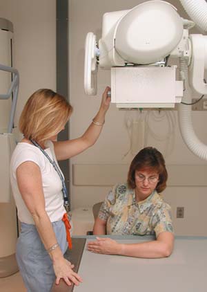
What does the equipment look like?
The equipment typically used for bone x-rays consists of an x-ray tube suspended over a table on which the patient lies. A drawer under the table holds the x-ray film or image recording plate. Sometimes the x-ray is taken with the patient standing upright, as in cases of knee x-rays.
Compact, portable x-ray machines can be taken to the patient in a hospital bed or the emergency room. The x-ray tube is connected to a flexible arm. The technologist extends the arm over the patient and places an x-ray film holder or image recording plate under the patient.
How does the procedure work?
X-rays are a form of radiation like light or radio waves. X-rays pass through most objects, including the body. The technologist carefully aims the x-ray beam at the area of interest. The machine produces a small burst of radiation that passes through your body. The radiation records an image on photographic film or a special detector.
Different parts of the body absorb the x-rays in varying degrees. Dense bone absorbs much of the radiation while soft tissue (muscle, fat, and organs) allow more of the x-rays to pass through them. As a result, bones appear white on the x-ray, soft tissue shows up in shades of gray, and air appears black.
Most x-ray images are electronically stored digital files. Your doctor can easily access these stored images to diagnose and manage your condition.
How is the procedure performed?
The technologist, an individual specially trained to perform radiology examinations, positions the patient on the x-ray table and places the x-ray film holder or digital recording plate under the table in the area of the body being imaged. When necessary, sandbags, pillows or other positioning devices will be used to help you maintain the proper position. A lead apron may be placed over your pelvic area or breasts when feasible to protect from radiation.
You must hold very still and may need to hold your breath for a few seconds while the technologist takes the x-ray. This helps reduce the possibility of a blurred image. The technologist will walk behind a wall or into the next room to activate the x-ray machine.
You may be repositioned for another view and the process is repeated. Two or three images (from different angles) will typically be taken.
An x-ray may also be taken of the unaffected limb, or of a child's growth plate (where new bone is forming), for comparison purposes.
When the examination is complete, the technologist may ask you to wait until the radiologist confirms they have all the necessary images.
A bone x-ray examination is usually completed within five to 10 minutes.
What will I experience during and after the procedure?
A bone x-ray examination itself is a painless procedure.
You may experience discomfort from the cool temperature in the examination room. You may also find holding still in a particular position and lying on the hard examination table uncomfortable, especially if you are injured. The technologist will assist you in finding the most comfortable position possible that still ensures x-ray image quality.
Who interprets the results and how do I get them?
A radiologist, a doctor trained to supervise and interpret radiology examinations, will analyze the images. The radiologist will send a signed report to your primary care or referring physician who will discuss the results with you.
You may need a follow-up exam. If so, your doctor will explain why. Sometimes a follow-up exam further evaluates a potential issue with more views or a special imaging technique. It may also see if there has been any change in an issue over time. Follow-up exams are often the best way to see if treatment is working or if a problem needs attention.
What are the benefits vs. risks?
Benefits
- Bone x-rays are the fastest and easiest way for a physician to view and assess bone injuries, including fractures, and joint abnormalities, such as arthritis.
- X-ray equipment is relatively inexpensive and widely available in emergency rooms, doctors’ offices, ambulatory care centers, nursing homes, and other locations. This makes it convenient for both patients and doctors.
- Because x-ray imaging is fast and easy, it is particularly useful in emergency diagnosis and treatment.
- No radiation stays in your body after an x-ray exam.
- X-rays usually have no side effects in the typical diagnostic range for this exam.
Risks
- There is always a slight chance of cancer from excessive exposure to radiation. However, given the small amount of radiation used in medical imaging, the benefit of an accurate diagnosis far outweighs the associated risk.
- The radiation dose for this procedure varies. See the Radiation Dose page for more information.
- Women should always tell their doctor and x-ray technologist if they are pregnant. See the Radiation Safety page for more information about pregnancy and x-rays.
A Word About Minimizing Radiation Exposure
Doctors take special care during x-ray exams to use the lowest radiation dose possible while producing the best images for evaluation. National and international radiology protection organizations continually review and update the technique standards radiology professionals use.
Modern x-ray systems minimize stray (scatter) radiation by using controlled x-ray beams and dose control methods. This ensures that the areas of your body not being imaged receive minimal radiation exposure.
What are the limitations of bone x-ray?
While x-ray images are among the clearest, most detailed views of bone, they provide little information about muscles, tendons or joints.
An MRI may be more useful in identifying bone and joint injuries (e.g., meniscal and ligament tears in the knee, rotator cuff and labrum tears in the shoulder) and in imaging of the spine (because both the bones and the spinal cord can be evaluated). MRI can also detect subtle or occult fractures or bone bruises (also called bone contusions or microfractures) not visible on x-ray images.
CT is being used widely to assess trauma patients in emergency departments. A CT scan can image complicated fractures, subtle fractures or dislocations. In elderly or patients with osteoporosis, a hip fracture may be clearly seen on a CT scan, while it may be barely seen, if at all, on a hip x-ray.
For suspected spine injury or other complicated injuries, 3-D reconstructed CT images can be made without additional radiation exposure to help the diagnosis and treatment of the individual patient's condition.
Ultrasound imaging, which uses sound waves instead of ionizing radiation to create diagnostic images, has also been useful for injuries around joints, and in evaluating the hips of children with congenital problems.
This page was reviewed on March 11, 2024