Esophageal Cancer Treatment
What is Esophageal Cancer?
Esophageal cancer occurs when cancer cells develop in the esophagus, a long, tube-like structure that connects the throat and the stomach. The esophagus, which carries swallowed food to the stomach, is part of the upper digestive system. The wall of the esophagus consists of several layers of tissue.
There are two main types of esophageal cancer, including:
- squamous cell carcinoma, in which cancer develops in the thin, flat cells (called squamous) that form the inner lining of the esophagus.
- adenocarcinoma, in which cancer develops in glandular cells in the lining of the esophagus.
In the early stages of esophageal cancer, there may be no symptoms. In more advanced cancers, symptoms may include:
- difficulty or pain when swallowing
- weight loss
- pain with swallowing or in the chest
- coughing and regurgitation
- hoarseness
- vomit blood
- tarry stool, or blood in stool
- indigestion and heartburn
What are my treatment options?
Treatment options include:
Surgery
Surgery to remove the cancer may be used alone for early stage disease or in combination with other therapies for advanced disease. If the cancer is a small tumor confined to the first layer of the lining in the esophagus, the surgeon may remove the tumor and a small amount of surrounding healthy tissue (called a margin).
In more advanced cancers, part of the esophagus may be removed. In an esophagectomy, the portion of the esophagus that contains the tumor along with nearby lymph nodes is removed and the remaining esophagus is re-connected to the stomach or part of the patient's gastrointestinal (GI) tract. In an esophagogastrectomy, the diseased part of the esophagus, nearby lymph nodes and part of the stomach are removed.
Endoscopic Treatments
Endoscopic treatments, which are used to treat early and pre-cancers of the esophagus and for pain relief (called palliative treatment), include:
- Endoscopic mucosal resection: In this procedure, a thin tube called an endoscope is inserted through the throat to the esophagus. The endoscope is equipped with a light, video camera and other instruments that are used to remove cancerous tissue in the esophagus.
Chemotherapy: This treatment uses chemical substances or drugs to kill cancer cells or stop them from dividing. Chemotherapy may be used before or after surgery for esophageal cancer. Chemotherapy is also used to help relieve symptoms when esophageal cancer has spread beyond the esophagus (metastasized).
Monoclonal Antibody Therapy (also called targeted therapy): A small number of esophageal cancers have too much of a protein called HER2 on the surface of their cells. A drug known as trastuzumab (Herceptin), is a monoclonal antibody that attaches to the HER2 protein on cancer cells and interferes with their ability to grow. This targeted therapy may be combined with chemotherapy.
Radiation therapy: This cancer treatment that uses high-energy x-rays or other types of radiation to kill cancer cells.
- Esophageal cancer patients may be treated with external beam therapy, in which beams of high-energy radiation generated by a machine outside the patient and directed at the tumor and cancerous lymph nodes. The types of radiation used to treat esophageal cancer patients include photons (x-rays and gamma rays) and protons through proton therapy.
Radiation therapy is usually combined with chemotherapy and surgery to treat patients with esophageal cancer and is often used for patients who are not candidates for surgery. For patients undergoing surgical treatment for esophageal cancer, radiation therapy may be used before surgery to help shrink the cancer (called neoadjuvant treatment) or after surgery, to destroy any remaining cancer cells (called adjuvant therapy). It may also be used to help manage the symptoms and complications of advanced disease, including pain and tumor growth that prohibits food from passing to the stomach.
What happens during radiation therapy?
Prior to beginning radiation therapy, some patients who cannot swallow may have a feeding tube inserted into their esophagus. This tube, called an esophageal stent, allows the esophagus to remain open.
The treatment process for external beam therapy begins with simulation and treatment planning.
The goal of simulation is to determine the patient's daily treatment position and to make devices that will help the patient maintain that position. Body molds, head masks, or other devices may be constructed to help the patient stay still during treatment and temporary skin marks or tattoos may also be applied to help precisely position the patient for each treatment session.
For treatment planning, computed tomography (CT), position emission tomography (PET) and magnetic resonance imaging (MRI) images will be obtained to map the location of the cancer and healthy tissues nearby. Using these images and sophisticated treatment planning tools, a team of professionals including a dosimetrist, radiation physicist and radiation oncologist generate a treatment plan that will deliver the appropriate radiation dose to the tumor while minimizing exposure to surrounding normal tissues.
After the simulation and planning have been completed, the treatment can begin.
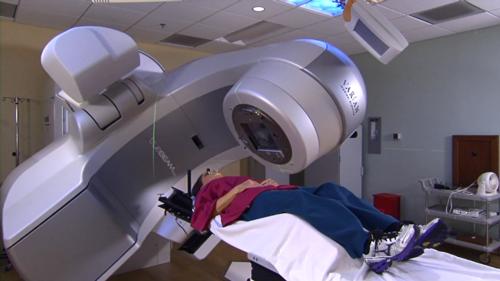
External beam therapy with high energy x-rays is often delivered from a machine called a linear accelerator. Various techniques are used to deliver EBT, including three-dimensional conformal radiation therapy (3D-CRT), intensity modulated radiation therapy (IMRT) and image-guided radiation therapy (IGRT).
- 3-D CRT more precisely conforms the radiation to the tumor, allowing a higher radiation dose to be safely delivered.
- IMRT uses special devices called collimators to regulate the intensity of the radiation beams, allowing different areas of a tumor and nearby tissues to receive different doses of radiation.
- IGRT is often used in conjunction with IMRT to deliver precise radiation doses to a malignant tumor or even specific areas within the tumor.
External beam radiation therapy can also be delivered using beams of a charged particle called a proton, which are typically generated by a machine called a cyclotron.
Before each radiation therapy session, the patient may be asked to change into a gown. The patient is then placed on the treatment couch in exactly the same position that was used for simulation using the immobilization devices. The therapist goes outside the room and turns on the linear accelerator from outside.
Patients undergo radiation therapy during a series of outpatient treatments over several weeks. The patient's diagnosis determines the total duration of treatment. Each treatment session lasts less than an hour, most of which is spent positioning the patient.
What are possible side effects of radiation therapy?
Radiation treatment can cause side effects. These problems may result from the treatment itself or from radiation damage to healthy cells in the treatment area.
The number and severity of side effects will depend on the type of radiation, dose, and body part under treatment. Talk to your doctor and/or nurse so they can help manage them.
Radiation can cause early and late side effects. Early side effects happen during or right after treatment. They are typically gone within a few weeks. Common early side effects include fatigue and skin problems. Skin in the treatment area may become sensitive, red, irritated, or swollen. Other changes include dryness, itching, peeling, and blistering.
Depending on the area being treated, other early side effects may include:
- hair loss in the treatment area
- mouth problems and difficulty swallowing
- eating and digestion problems
- diarrhea
- nausea and vomiting
- headaches
- soreness and swelling in the treatment area
- urinary and bladder changes
Late side effects may occur months or years following treatment. While they are often permanent, they are rare. They include:
- brain changes
- spinal cord changes
- lung changes
- kidney changes
- colon and rectal changes
- infertility
- joint changes
- lymphedema
- mouth changes
- secondary cancer
There is a slight risk of developing cancer from radiation therapy. After treatment, your radiation oncologist will regularly check for complications and recurrent or new cancers.
Individuals undergoing radiation therapy may experience early or acute side effects during the course of treatment. They may also experience chronic or late side effects months or even years after they have completed treatment. Side effects vary from individual to individual and may depend on one's general health, the area of the body undergoing treatment, the daily radiation dose, the total radiation dose given during treatment and other treatments (such as chemotherapy) they may be undergoing at the same time.
Patients undergoing external beam radiation treatment typically experience difficult and painful swallowing soon after starting treatment. This occurs as a result of the radiation killing the cells in the lining of the esophagus, which causes a reaction similar to a sunburn. It is important for patients to drink plenty of water and to meet their nutritional needs during this time. When patients cannot get adequate nutrition and hydration, a feeding tube may need to be inserted before or during radiation treatment until the patient regains the ability to swallow. Once treatment is completed, the pain usually subsides within a few weeks.
Other common side effects of radiation therapy include:
- fatigue
- skin changes, including dryness, itching, peeling, and blistering
- diarrhea
- dry mouth and other mouth problems
- extreme fatigue
- nausea (especially if the abdomen is treated)
- inflammation in the treated area, such as difficulty with swallowing, a cough or feeling short of breath as a result of radiation to the chest
- pain with or difficulty swallowing
- decreased blood cell counts, which can lead to increased fatigue, an increased risk of infection and bruising
- loss of hair in the treatment area
Most of these side effects go away within two months after radiation therapy is finished.
Late or long-term side effects may occur six or more months after radiation therapy is completed. Whether or not a patient experiences late side effects depends on the area of the body treated with radiation therapy, other aspects of their cancer treatment and individual risk factors. Radiation therapy for esophageal cancer may cause a stricture (narrowing) in the esophagus, which may require further treatment. Other possible long-term side effects include:
- fibrosis (the replacement of normal tissue with scar tissue, leading to restricted movement of the affected area)
- damage to the bowels, causing diarrhea and bleeding
- infertility
- heart or lung damage from radiation therapy to the chest, possibly leading to problems breathing and shortness of breath.
- thyroid problems or irritation of the esophagus from radiation to the neck
- headaches and problems with memory loss, personality changes and trouble concentrating from radiation to the head
- the development of another cancer later in life, called secondary cancer
Patients should talk to their doctor before and during treatment about the side effects they can expect and ways to minimize them.
Are there any new developments in treating my disease?
- New imaging modalities, such as endoscopic ultrasound, PET-CT, and probe-based confocal laser endomicroscopy, a technique that provides real-time microscopic views of the esophagus, are improving the detection of esophageal cancer.
- Advances in surgical resection techniques for esophageal cancer are offering improved patient outcomes. The use of minimally invasive techniques, such as the endoscopic mucosal resection, is improving survival rates for the disease.
This page was reviewed on September 05, 2018