Lymphoscintigraphy
Lymphoscintigraphy helps evaluate your body's lymphatic system for disease using small amounts of radioactive materials called radiotracers that are typically injected into the bloodstream, inhaled, swallowed, or in the case of lymphoscintigraphy, injected into the skin. The radiotracer travels through the area being examined and gives off energy in the form of gamma rays which are detected by a special camera and a computer to create images of the inside of your body. Because it is able to pinpoint molecular activity within the body, lymphoscintigraphy offers the potential to identify lymphatic disease in its earliest stages.
Tell your doctor if there's a possibility you are pregnant or if you are breastfeeding. Discuss any recent illnesses, medical conditions, allergies and medications you're taking, including vitamins and herbal supplements. Your doctor will instruct you on how to prepare. Leave jewelry at home and wear loose, comfortable clothing. You may be asked to wear a gown.
- What is Lymphoscintigraphy?
- What are some common uses of the procedure?
- How should I prepare?
- What does the equipment look like?
- How does the procedure work?
- How is the procedure performed?
- What will I experience during and after the procedure?
- Who interprets the results and how do I get them?
- What are the benefits vs. risks?
- What are the limitations of Lymphoscintigraphy?
What is Lymphoscintigraphy?
Lymphoscintigraphy is a special type of nuclear medicine imaging that provides pictures called scintigrams of the lymphatic system.
Nuclear medicine uses small amounts of radioactive material called radiotracers. Doctors use nuclear medicine to diagnose, evaluate, and treat various diseases. These include cancer, heart disease, gastrointestinal, endocrine, or neurological disorders, and other conditions. Nuclear medicine exams pinpoint molecular activity. This gives them the potential to find disease in its earliest stages. They can also show whether you are responding to treatment.
Nuclear medicine is noninvasive. Except for intravenous injections, it is usually painless. These tests use radioactive materials called radiopharmaceuticals or radiotracers to help diagnose and assess medical conditions.
Radiotracers are molecules linked to, or "labeled" with, a small amount of radioactive material. They accumulate in tumors or regions of inflammation. They can also bind to specific proteins in the body. The most common radiotracer is F-18 fluorodeoxyglucose (FDG), a molecule similar to glucose. Cancer cells are more metabolically active and may absorb glucose at a higher rate. This higher rate can be seen on PET scans. This allows your doctor to detect disease before it may be seen on other imaging tests. FDG is just one of many radiotracers in use or in development.
You will usually receive the radiotracer in an injection. Or you may swallow it or inhale it as a gas, depending on the exam. It accumulates in the area under examination. A special camera detects gamma ray emissions from the radiotracer. The camera and a computer produce pictures and supply molecular information.
The lymphatic system is a network of small channels similar to blood vessels that circulate the fluid (called lymph) and cells (lymphocytes) of the immune system throughout the body. Lymph nodes, which act like a filter for foreign bodies such as germs, viruses and pollen, are located along this network.
What are some common uses of the procedure?
Physicians perform lymphoscintigraphy to:
- identify the sentinel lymph node, or the first node to receive the lymph drainage from a tumor.
- plan a biopsy or surgery that will help assess the stage of cancer and create a treatment plan.
- identify points of blockage in the lymphatic system, such as lymph flow in an arm or leg, or lymphedema.
How should I prepare?
You may wear a gown during the exam or be allowed to wear your own clothing.
Women should always tell their doctor and technologist if they are pregnant or breastfeeding. See the Radiation Safety page for more information about pregnancy and breastfeeding related to nuclear medicine imaging.
Tell the doctor and your exam technologist about any medications you are taking, including vitamins and herbal supplements. List any allergies, recent illnesses, and other medical conditions.
Leave jewelry and accessories at home or remove them prior to the exam. These objects may interfere with the procedure.
Your doctor will tell you how to prepare for your specific exam.
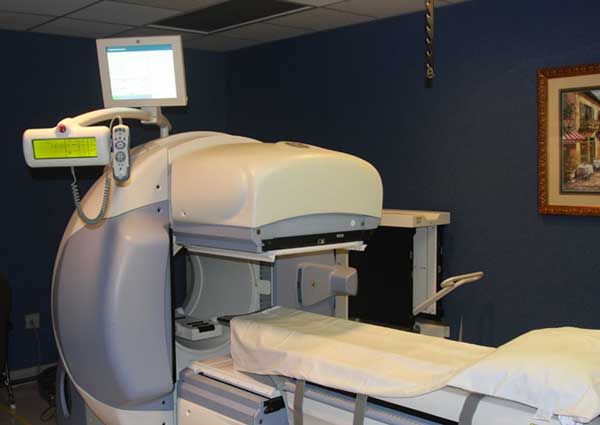
What does the equipment look like?
Nuclear medicine uses a special gamma camera and single-photon emission-computed tomography (SPECT) imaging techniques.
The gamma camera records the energy emissions from the radiotracer in your body and converts it into an image. The gamma camera itself does not emit any radiation. It has radiation detectors called gamma camera heads. These are encased in metal and plastic, often shaped like a box, and attached to a round, donut-shaped gantry. The patient lies on an exam table that slides in between two parallel gamma camera heads, above and beneath the patient. Sometimes, the doctor will orient the gamma camera heads at a 90-degree angle over the patient's body.
In SPECT, the gamma camera heads rotate around the patient's body to produce detailed, three-dimensional images.
A computer creates the images using the data from the gamma camera.
A probe is a small hand-held device resembling a microphone. It measures the amount of radiotracer in an area of your body.
How does the procedure work?
Ordinary x-ray exams pass x-rays through the body to create an image. Nuclear medicine uses radioactive materials called radiopharmaceuticals or radiotracers. Your doctor typically injects this material into your bloodstream. Or you may swallow it or inhale it as a gas. The material accumulates in the area under examination, where it gives off gamma rays. Special cameras detect this energy and, with the help of a computer, create pictures that detail how your organs and tissues look and function.
How is the procedure performed?
Doctors perform nuclear medicine exams on outpatients and hospitalized patients.
You will lie on an exam table. If necessary, a nurse or technologist will insert an intravenous (IV) catheter into a vein in your hand or arm.
The radiotracer will be injected just beneath the skin, or sometimes deeper, using a very small needle.
Immediately after the injection, the gamma camera will take a series of images of the area of the body being studied.
When imaging begins, the camera or scanner will take a series of images. The camera may rotate around you or stay in one position. You may need to change positions in between images. While the camera is taking pictures, you will need to remain still for brief periods. In some cases, the camera may move very close to your body. This is necessary to obtain the best quality images. Tell the technologist if you have a fear of closed spaces before your exam begins.
The type of study you are having will determine the location of your injection and the number of scans performed.
- Melanoma cancer patients — Two to five doses of radiotracer are injected into the skin or other tissue surrounding the site of the melanoma. Images may be taken of the arms and underarms, legs and groins, or head, neck and chest, or other areas, depending on the site of the melanoma. Your skin will be marked to show where your lymph nodes are located. Imaging for this procedure usually takes about one to two hours, but may take up to three to four hours.
- Breast cancer — The radiotracer may be injected in multiple sites near the tumor and/or around the areola, or nipple. The breast, chest and underarm regions will be imaged. Imaging usually is completed within 30 minutes to one hour, but may take up to two or more hours.
- Leg or arm swelling (edema) — The radiotracer is injected between the first and second fingers or toes of each hand or foot. Both the swollen and healthy arm or leg will be imaged so that the two sides can be compared. Depending on the degree of lymphatic obstruction and the cause, imaging may take 30 minutes to several hours.
For some procedures, you may also be asked to exercise lightly for about 10 minutes—walking for leg exams or doing handgrip or lifting exercises for arm exams. Additional images are taken once you complete these exercises.
After the exam, you may need to wait until the technologist determines if more images are needed. Sometimes, the technologist takes more images to clarify or better visualize certain areas or structures. The need for more images does not necessarily mean there was a problem with the exam or that something is abnormal. It should not cause you concern.
If you have an intravenous (IV) line for the procedure, your technologist will usually remove it. The technologist will leave it in place if you are to have another procedure that same day that requires an IV line.
What will I experience during and after the procedure?
Except for intravenous injections, most nuclear medicine procedures are painless. Reports of significant discomfort or side effects are rare.
No anesthesia is needed for a scintigram unless a lymph node biopsy is performed in the operating room following the procedure.
You will feel a slight pin prick when the technologist inserts the needle into your vein for the intravenous line. You may feel a cold sensation moving up your arm during the radiotracer injection. Generally, there are no other side effects.
For lymphoscintigraphy or sentinel node studies, the radiotracer is not injected intravenously, but rather near the tumor site.
It is important to remain still during the exam. Nuclear imaging causes no pain. However, having to remain still or in one position for long periods may cause discomfort.
Unless your doctor tells you otherwise, you may resume your normal activities after your exam. A technologist, nurse, or doctor will provide you with any necessary special instructions before you leave.
The small amount of radiotracer in your body will lose its radioactivity over time through the natural process of radioactive decay. It may also pass out of your body through your urine or stool during the first few hours or days after the test. Drink plenty of water to help flush the material out of your body.
Who interprets the results and how do I get them?
A radiologist or other doctor specially trained in nuclear medicine will interpret the images and send a report to your referring physician.
What are the benefits vs. risks?
Benefits
- This nuclear medicine test has essentially replaced the more complex procedure formerly used to assess the lymphatic system as well as to determine the spread of cancer to lymph nodes (lymphangiography).
- Lymphoscinitigraphy allows for a less extensive surgery to be performed which has fewer side effects and a lower morbidity rate compared to more radical surgery (axillary lymph node dissection).
- Nuclear medicine exams provide unique information that is often unattainable using other imaging procedures. This information may include details on the function and anatomy of body structures.
- Nuclear medicine supplies the most useful diagnostic or treatment information for many diseases.
- A nuclear medicine scan is less expensive and may yield more precise information than exploratory surgery.
Risks
- Because nuclear medicine exams use only a small dose of radiotracer, they have a relatively low radiation exposure. This is acceptable for diagnostic exams. Thus, the potential benefits of an exam outweigh the very low radiation risk.
- Doctors have been using nuclear medicine diagnostic procedures for more than six decades. There are no known long-term adverse effects from such low-dose exposure.
- Your doctor always weighs the benefits of nuclear medicine treatment against any risks. Your doctor will discuss the significant risks prior to treatment and give you an opportunity to ask questions.
- Allergic reactions to radiotracers are extremely rare and usually mild. Always tell the nuclear medicine personnel about any allergies you may have. Describe any problems you may have had during previous nuclear medicine exams.
- The radiotracer injection may cause slight pain and redness. This should rapidly resolve.
- Women should always tell their doctor and radiology technologist if there is any possibility that they are pregnant, or they are breastfeeding. See the Radiation Safety page for more information about pregnancy, breastfeeding and nuclear medicine exams.
What are the limitations of Lymphoscintigraphy?
Nuclear medicine procedures can be time consuming. It can take several hours to days for the radiotracer to accumulate in the area of interest. Plus, imaging may take up to several hours to perform. In some cases, newer equipment can substantially shorten the procedure time.
The image resolution of nuclear medicine images may not be as high as that of CT or MRI. However, nuclear medicine scans are more sensitive for a variety of indications. The functional information they yield is often unobtainable using other imaging techniques.
Additional Information and Resources
RTAnswers.org: Radiation Therapy for Lymphomas
This page was reviewed on April 15, 2022