MRCP (MR Cholangiopancreatography)
Magnetic resonance cholangiopancreatography or MRCP uses a powerful magnetic field, radio waves and a computer to evaluate the liver, gallbladder, bile ducts, pancreas and pancreatic duct for disease. It is noninvasive and does not use ionizing radiation.
Tell your doctor about any health problems, recent surgeries or allergies and whether there's a possibility you are pregnant. The magnetic field is not harmful, but it may cause some medical devices to malfunction. Most orthopedic implants pose no risk, but you should always tell the technologist if you have any implanted devices or metal in your body. Guidelines about eating and drinking before your exam vary between facilities. Unless you are told otherwise, take your regular medications as usual. Leave jewelry at home and wear loose, comfortable clothing. You may be asked to wear a gown. If you have claustrophobia or anxiety, you may want to ask your doctor for a mild sedative prior to the exam.
- What is MRCP?
- What are some common uses of the procedure?
- How should I prepare?
- What does the equipment look like?
- How does the procedure work?
- How is the procedure performed?
- What will I experience during and after the procedure?
- Who interprets the results and how do I get them?
- What are the benefits vs. risks?
- What are the limitations of MRCP?
What is MRCP?
Magnetic resonance cholangiopancreatography (MRCP) is a special type of magnetic resonance imaging (MRI) exam that produces detailed images of the hepatobiliary and pancreatic systems, including the liver, gallbladder, bile ducts, pancreas and pancreatic duct.
Magnetic resonance imaging (MRI) is a noninvasive test radiologists use to diagnose medical conditions.
MRI uses a powerful magnetic field, radiofrequency pulses, and a computer to produce detailed pictures of internal body structures. MRI does not use radiation (x-rays).
Detailed MR images allow radiologists to examine the body and detect disease.
What are some common uses of the procedure?
Physicians use MRCP to:
- examine diseases of the liver, gallbladder, bile ducts, pancreas and pancreatic duct. These may include tumors, stones, inflammation or infection.
- evaluate patients with pancreatitis to detect the underlying cause. In patients with pancreatitis, an MRCP may be performed using a medication called Secretin to assess for long term scarring and to determine the amount of healthy pancreatic function and secretions.
- help to diagnose unexplained abdominal pain.
- provide a noninvasive alternative to endoscopic retrograde cholangiopancreatography (ERCP). ERCP is a diagnostic procedure that combines endoscopy, which uses an illuminated optical instrument to examine inside the body, with iodinated contrast injection and x-ray images. ERCP is an invasive procedure that evaluates the bile ducts and/or the pancreatic duct.
How should I prepare?
You may be asked to wear a gown during the exam or you may be allowed to wear your own clothing if it is loose-fitting and has no metal fasteners.
Guidelines about eating and drinking before an MRI exam vary at different facilities. Usually, you will be instructed not to eat or drink anything for several hours before your procedure.
Because your procedure may require use of contrast material that is swallowed or injected into your bloodstream, the radiologist or technologist may ask if you have allergies of any kind, including allergies to food or drugs, hay fever, hives or allergic asthma. However, the contrast material used for an MRI exam is based on gadolinium and does not contain iodine. A gadolinium contrast agent is less likely to cause an allergic reaction compared to the iodinated contrast agents used in CT scanning.
The radiologist should also know if you have any serious health problems and what surgeries you have undergone. Some conditions, such as kidney disease, may prevent you from having an MRI with contrast material.
Women should always tell their doctor and technologist if they are pregnant. MRI has been used since the 1980s with no reports of any ill effects on pregnant women or their unborn babies. However, the baby will be in a strong magnetic field. Therefore, pregnant women should not have an MRI in the first trimester unless the benefit of the exam clearly outweighs any potential risks. Pregnant women should not receive gadolinium contrast unless absolutely necessary. See the MRI Safety During Pregnancy page for more information about pregnancy and MRI.
If you have claustrophobia (fear of enclosed spaces) or anxiety, ask your doctor to prescribe a mild sedative prior to the date of your exam.
Leave all jewelry and other accessories at home or remove them prior to the MRI scan. Metal and electronic items are not allowed in the exam room. They can interfere with the magnetic field of the MRI unit, cause burns, or become harmful projectiles. These items include:
- jewelry, watches, credit cards, and hearing aids, all of which can be damaged
- pins, hairpins, metal zippers, and similar metallic items, which can distort MRI images
- removable dental work
- pens, pocketknives, and eyeglasses
- body piercings
- mobile phones, electronic watches, and tracking devices.
In most cases, an MRI exam is safe for patients with metal implants, except for a few types. People with the following implants may not be scanned and should not enter the MRI scanning area without first being evaluated for safety:
- some cochlear (ear) implants
- some types of clips used for brain aneurysms
- some types of metal coils placed within blood vessels
- some older cardiac defibrillators and pacemakers
- vagal nerve stimulators
Tell the technologist if you have medical or electronic devices in your body. These devices may interfere with the exam or pose a risk. Many implanted devices will have a pamphlet explaining the MRI risks for that device. If you have the pamphlet, bring it to the attention of the scheduler before the exam. MRI cannot be performed without confirmation and documentation of the type of implant and MRI compatibility. You should also bring any pamphlet to your exam in case the radiologist or technologist has any questions.
If there is any question, an x-ray can detect and identify any metal objects. Metal objects used in orthopedic surgery generally pose no risk during MRI. However, a recently placed artificial joint may require the use of a different imaging exam.
Tell the technologist or radiologist about any shrapnel, bullets, or other metal that may be in your body. Foreign bodies near and especially lodged in the eyes are very important because they may move or heat up during the scan and cause blindness. Dyes used in tattoos may contain iron and could heat up during an MRI scan. This is rare. The magnetic field will usually not affect tooth fillings, braces, eyeshadows, and other cosmetics. However, these items may distort images of the facial area or brain. Tell the technologist or radiologist about them.
Your child may need to be sedated in order to hold still adequately during the procedure. If this is the case, you will be given instructions for your child about not eating or drinking several hours prior to sedation and the examination. For the safety of your child during the sedation, it is important that you fully understand and follow any instructions that have been given. After the procedure there will be a recovery period from the sedation. Your child will be discharged when the nurses and physicians believe he/she is sufficiently awake to be safely sent home.
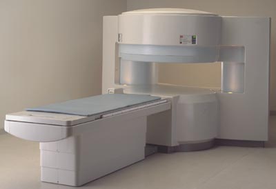
What does the equipment look like?
The traditional MRI unit is a large cylinder-shaped tube surrounded by a circular magnet. You will lie on a table that slides into a tunnel towards the center of the magnet.
Some MRI units, called short-bore systems, are designed so that the magnet does not completely surround you. Some newer MRI machines have a larger diameter bore, which can be more comfortable for larger patients or those with claustrophobia. "Open" MRI units are open on the sides. They are especially helpful for examining larger patients or those with claustrophobia. Open MRI units can provide high quality images for many types of exams. Open MRI may not be used for certain exams. For more information, consult your radiologist.
How does the procedure work?
Unlike x-ray and computed tomography (CT) exams, MRI does not use radiation. Instead, radio waves re-align hydrogen atoms that naturally exist within the body. This does not cause any chemical changes in the tissues. As the hydrogen atoms return to their usual alignment, they emit different amounts of energy depending on the type of tissue they are in. The scanner captures this energy and creates a picture using this information.
In most MRI units, the magnetic field is produced by passing an electric current through wire coils. Other coils are inside the machine and, in some cases, are placed around the part of the body being imaged. These coils send and receive radio waves, producing signals that are detected by the machine. The electric current does not come into contact with the patient.
A computer processes the signals and creates a series of images, each of which shows a thin slice of the body. The radiologist can study these images and visualize the body from different angles.
MRI is often able to tell the difference between diseased tissue and normal tissue better than x-ray, CT, and ultrasound.
How is the procedure performed?
MRI exams may be done on an outpatient basis.
The technologist will position you on the moveable exam table. They may use straps and bolsters to help you stay still and maintain your position.
The technologist may place devices that contain coils capable of sending and receiving radio waves around or next to the area of the body under examination.
MRI exams generally include multiple runs (sequences), some of which may last several minutes. Each run will create a different set of noises.
If your exam uses a contrast material, a doctor, nurse, or technologist will insert an intravenous catheter (IV line) into a vein in your hand or arm. They will use this IV to inject the contrast material.
You will be placed into the magnet of the MRI unit. The technologist will perform the exam while working at a computer outside of the room. You will be able to talk to the technologist via an intercom.
If your exam uses a contrast material, the technologist will inject it into the intravenous line (IV) after an initial series of scans. They will take more images during or following the injection.
The actual MRCP exam takes approximately 10-15 minutes, but it is often performed with a standard MRI of the abdomen, which may last approximately 30 minutes and involves the use of contrast material. In this case, the entire examination is usually completed within 45 minutes.
What will I experience during and after the procedure?
Most MRI exams are painless. However, some patients find it uncomfortable to remain still. Others may feel closed-in (claustrophobic) while in the MRI scanner. The scanner can be noisy.
If contrast material is used, there may be brief discomfort during initial placement of the intravenous catheter line. The oral contrast used at some institutions may have an unpleasant taste and cause temporary fullness, but most patients tolerate it well.
It is normal for the area of your body being imaged to feel slightly warm. If it bothers you, tell the radiologist or technologist. It is important that you remain perfectly still while the images are being taken. This is typically only a few seconds to a few minutes at a time. You will know when images are being recorded because you will hear and feel loud tapping or thumping sounds. The coils that generate the radio waves make these sounds when they are activated. You will be provided with earplugs or headphones to reduce the noise made by the scanner. You may be able to relax between imaging sequences. However, you will need to keep the same position as much as possible without moving.
You will usually be alone in the exam room. However, the technologist will be able to see, hear, and speak with you at all times using a two-way intercom. They will give you a “squeeze-ball” that alerts the technologist that you need attention right away. Many facilities allow a friend or parent to stay in the room if they have also been screened for safety.
Children will be given appropriately sized earplugs or headphones during the exam. Music may be played through the headphones to help pass the time. MRI scanners are air-conditioned and well-lit.
In some cases, IV injection of contrast material may be given before the images are obtained. The IV needle may cause you some discomfort and you may experience some bruising. There is also a very small chance of skin irritation at the site of the IV tube insertion. Some patients may have a temporary metallic taste in their mouth after the contrast injection.
If you do not require sedation, no recovery period is necessary. You may resume your usual activities and normal diet immediately after the exam. On very rare occasions, a few patients experience side effects from the contrast material. These may include nausea, headache, and pain at the site of injection. It is very rare that patients experience hives, itchy eyes, or other allergic reactions to the contrast material. If you have allergic symptoms, tell the technologist. A radiologist or other doctor will be available for immediate assistance.
Who interprets the results and how do I get them?
A radiologist, a doctor trained to supervise and interpret radiology exams, will analyze the images. The radiologist will send a signed report to your primary care or referring physician, who will share the results with you. Most imaging facilities use an electronic medical record. Frequently, there will be a website or mobile application to provide patient access to the electronic medical record and you may be able to access your imaging report digitally but usually not the images. You can ask the technologist if this is available at the imaging facility.
What are the benefits vs. risks?
Benefits
- MRI is a noninvasive imaging technique that does not involve exposure to radiation.
- MRI can provide detailed images of the soft-tissue structures of the body—such as the heart, liver, pancreas and many other organs. This detail makes MRI an invaluable tool in early diagnosis and evaluation of cancer.
- MRI has proven valuable in diagnosing a broad range of conditions, including heart and vascular disease, stroke, and joint and musculoskeletal disorders.
- MRI can help physicians evaluate both the structure of an organ and how it is working.
- MRI can detect abnormalities that might be obscured by bone with other imaging methods.
- The MRI gadolinium contrast material is less likely to cause an allergic reaction than the iodine-based contrast materials used for x-rays and CT scanning.
- MRCP can produce images comparable to those obtained by a more invasive exam called endoscopic retrograde cholangiopancreatography (ERCP) without its associated risks including pancreatitis, or inflammation of the pancreas, perforation of pancreatic and bile ducts and bowel, and the risks for intravenous sedation required for ERCP.
Risks
- The MRI exam poses almost no risk to the average patient when appropriate safety guidelines are followed.
- If sedation is used, there is a risk of using too much. However, your vital signs will be monitored to minimize this risk.
- The strong magnetic field is not harmful to you. However, it may cause implanted medical devices to malfunction or distort the images.
- Nephrogenic systemic fibrosis is a recognized complication related to injection of gadolinium contrast. It is exceptionally rare with the use of newer gadolinium contrast agents. It usually occurs in patients with serious kidney disease. Your doctor will carefully assess your kidney function before considering a contrast injection.
- There is a very slight risk of an allergic reaction if your exam uses contrast material. Such reactions are usually mild and controlled by medication. If you have an allergic reaction, a doctor will be available for immediate assistance.
- Although there are no known health effects, evidence has shown that very small amounts of gadolinium can remain in the body, particularly the brain, after multiple MRI exams. This is most likely to occur in patients receiving multiple MRI exams over their lifetime for monitoring chronic or high-risk health conditions. The contrast agent is mostly eliminated from the body through the kidneys. If you are a patient in this category, consult with your doctor about the possibility of gadolinium retention, as this effect varies from patient to patient.
- Reaction to the oral contrast given at some institutions for MRCP is very rare. Also, the oral contrast used at some institutions may have an unpleasant taste and cause temporary fullness, but most patients tolerate it well.
- IV contrast manufacturers indicate mothers should not breastfeed their babies for 24-48 hours after contrast material is given. However, the most recent American College of Radiology (ACR) Manual on Contrast Media reports that studies show the amount of contrast absorbed by the infant during breastfeeding is extremely low. Guidelines for diagnostic imaging during pregnancy and lactation from the American College of Obstetricians and Gynecologists advise that breastfeeding should not be interrupted after gadolinium administration. For further information please consult the ACR Manual on Contrast Media and its references.
What are the limitations of MRCP?
High-quality images depend on your ability to remain perfectly still and follow breath-holding instructions while the images are being recorded. If you are anxious, confused or in severe pain, you may find it difficult to lie still during imaging.
A person who is very large may not fit into certain types of MRI machines. There are weight limits on the scanners.
Implants and other metallic objects can make it difficult to obtain clear images. Patient movement can have the same effect.
A very irregular heartbeat may affect the quality of images. This is because some techniques time the imaging based on the electrical activity of the heart.
MRI is generally not recommended for seriously injured patients. However, this decision is based on clinical judgment. This is because traction devices and life support equipment may distort the MR images. As a result, they must be kept away from the area to be imaged. Some trauma patients, however, may need MRI.
Present data show no convincing evidence that non contrast MRI harms the fetus of a pregnant woman. However, if the need for the exam is not time sensitive your doctor may delay the exam until after delivery. MRI gadolinium contrast agents are generally avoided during pregnancy except in very specific circumstances. Your doctor will discuss the benefits and risks of any MRI procedure with you. Doctors may perform MRI after the first trimester to assess the fetus for findings that are not fully evaluated by ultrasound.
This page was reviewed on April 01, 2024