Crohn’s Disease
Crohn's disease causes inflammation and irritation in the gastrointestinal tract, most commonly in the end part of the small intestine (ileum). Unexplained abdominal pain, diarrhea, rectal bleeding, weight loss and fever are all symptoms of Crohn's disease and are very similar to those of ulcerative colitis.
Your doctor will likely perform a physical exam to evaluate your condition and use blood and stool tests to look for signs of bleeding and rule out other underlying causes, such as infection. You may also undergo flexible sigmoidoscopy, colonoscopy, body CT, body MRI, MR enterography, upper GI, small bowel follow-through, or lower GI to confirm a diagnosis. No cure exists for Crohn's disease, but your doctor may prescribe medications, surgery or a special diet to help manage your condition.
What is Crohn's disease?
Crohn's disease is a type of inflammatory bowel disease (IBD) that causes inflammation and irritation in the gastrointestinal (GI) tract. It occurs most commonly in the end part of the small intestine, called the ileum.
The most common symptoms of Crohn's disease are abdominal pain, often in the lower right abdomen area, and diarrhea. Other symptoms include:
- Rectal bleeding
- Weight loss
- Fever
Symptoms of Crohn's disease result from an inappropriate activation of the immune system. The causes are unknown, but there is evidence that genetics plays a significant role.
The symptoms of Crohn's disease are very similar to those of ulcerative colitis, except that Crohn's disease can happen anywhere along the digestive tract, from the mouth to the anus, while ulcerative colitis is restricted to the large bowel (colon). Crohn's disease may lead to deep ulcers in the intestinal tract, giving a "cobblestone" appearance. Inflammation can lead to a buildup of scar tissue over time, slowing the movement of food through the intestine and causing severe cramps.
Crohn's disease can affect both children and adults. Approximately 20 percent of those diagnosed have symptoms before age 20.
How is Crohn's disease diagnosed and evaluated?
Your primary doctor will begin by asking about your medical history and symptoms. You will also undergo a physical exam.
Blood and stool tests can be used to look for signs of bleeding and rule out other causes of GI diseases, such as infection.
Other common tests for Crohn's disease include:
- Flexible sigmoidoscopy, performed by inserting a sigmoidoscope (a flexible tube with a tiny camera on the end) into the rectum to view the lower colon and rectum.
- Colonoscopy—the most commonly used test for Crohn's disease—uses a lighted instrument called a colonoscope to view the rectum and the entire colon. The last part of the small intestine, the ileum, can also sometimes be seen.
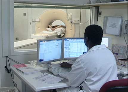
- Body CT scan, a special type of x-ray imaging that creates detailed pictures of your pelvis, abdomen or chest. You may receive an injection of contrast material so that the GI tract and abdominal organs show up more clearly in the pictures. CT enterography is a special CT scan that is better able to visualize the small intestine. This test has replaced barium enema and small bowel follow-through in many medical centers.
For more information about CT exams performed on children, visit the Pediatric CT page.
- Body MRI is a large machine that uses a strong magnetic field and radio waves to create detailed pictures of organs and other soft tissues in the abdomen.
- MR enterography is a special type of MRI performed with a contrast material to produce detailed images of the small intestine.
Infants and young children often require sedation or anesthesia to complete an MRI exam without moving. This depends on the child's age, intellectual development, and the type of exam. Sedation can be provided at many facilities. A specialist in pediatric sedation or anesthesia should be available during the exam for your child's safety. You will be told how to prepare your child.
Some facilities may have personnel who work with children to help avoid the need for sedation or anesthesia. They may prepare children by showing them a model MRI scanner and playing the noises they might hear during the exam. They also answer any questions and explain the procedure to relieve anxiety. Some facilities also provide goggles or headsets so the child can watch a movie during the exam. This helps the child stay still and allows for good quality images.
- Upper Gastrointestinal Tract Radiography, also called an upper GI series, is an x-ray examination of the esophagus, stomach and first part of the small intestine (also known as the duodenum). Images are produced using a special form of x-ray called fluoroscopy and an orally ingested contrast material such as barium.
- Small Bowel Follow-Through, in which an oral contrast material such as barium is ingested and the lower parts of the small intestine (jejunum and ileum) are viewed using abdominal radiographs (x-rays) and fluoroscopy.
- Lower Gastrointestinal Tract Radiography, also called a lower GI or barium enema, is an x-ray examination of the large intestine, also known as the colon. This examination evaluates the right or ascending colon, the transverse colon, the left or descending colon, the sigmoid colon and the rectum. The appendix and the very end of the small intestine may also sometimes be seen.
Infants and children may undergo upper and lower GI radiography. Usually, there is no special preparation, but your doctor will give you detailed instructions if needed. The use of barium and the means of capturing x-ray images are similar to that described for adults.
How is Crohn's disease treated?
Though no cure exists for Crohn's disease, treatment can improve symptoms, control inflammation and correct nutritional deficiencies. Among the common treatments options are:
- Medications: Anti-inflammation medications and antibiotics can help control inflammation, suppress the immune system and treat bacterial overgrowth in the small intestine.
- Proctocolectomy: This surgical procedure involves removal of the rectum and part of the colon or the entire colon. After removing the colon, the surgeon will perform an ileostomy, a procedure in which the small intestine is joined to an opening in the lower abdomen. A pouch is then attached to the opening and worn outside the body to collect stool. Sometimes, the small bowel can be attached to the anus so that a pouch is not needed.
- Intestinal resection surgery: The diseased section of intestine is removed and the ends of the healthy sections are connected.
- Nutrition supplementation: High-calorie liquid formulas or intravenous nutrition are sometimes given to Crohn's disease patients to give the inflamed intestine a respite from solid food.
Which test, procedure or treatment is best for me?
This page was reviewed on December 06, 2022