CT Enterography
Computed tomography (CT) enterography uses special x-ray equipment and an injection of contrast material after the ingestion of liquid to produce detailed images of the small intestine and structures within the abdomen and pelvis. It's often used to identify and locate problems within the bowel, such as inflammation, bleeding, obstructions and Crohn's disease. CT scanning is fast, painless, noninvasive and accurate. CT enterography is better able to visualize the entire thickness of the bowel wall when compared to other small intestine imaging procedures.
Tell your doctor if there's a possibility you are pregnant and discuss any recent illnesses, medical conditions, medications you're taking, and allergies. You will be instructed not to eat or drink anything for a few hours beforehand. If you have a known allergy to contrast material, your doctor may prescribe medications to reduce the risk of an allergic reaction. These medications must be taken 12 hours prior to your exam. Leave jewelry at home and wear loose, comfortable clothing. You may be asked to wear a gown.
- What is CT Enterography?
- What are some common uses of the procedure?
- How should I prepare?
- What does the equipment look like?
- How does the procedure work?
- How is the procedure performed?
- What will I experience during and after the procedure?
- Who interprets the results and how do I get them?
- What are the benefits vs. risks?
- What are the limitations of CT Enterography?
What is CT Enterography?
CT enterography is a special type of computed tomography (CT) imaging performed with intravenous contrast material after the ingestion of liquid that helps produce high resolution images of the small intestine in addition to the other structures in the abdomen and pelvis.
Computed tomography, more commonly known as a CT or CAT scan, is a diagnostic medical imaging test. Like traditional x-rays, it produces multiple images or pictures of the inside of the body.
A CT scan generates images that can be reformatted in multiple planes. It can even generate three-dimensional images. Your doctor can review these images on a computer monitor, print them on film or via a 3D printer, or transfer them to a CD or DVD.
CT images of internal organs, bones, soft tissue, and blood vessels provide greater detail than traditional x-rays. This is especially true for soft tissues and blood vessels.
What are some common uses of the procedure?
Physicians use CT enterography to identify and locate:
- small bowel inflammation
- bleeding sources within the small bowel
- small bowel tumors
- abscesses and fistulas
- bowel obstruction.
CT enterography is also used to diagnose Crohn's disease, and determine its location, severity and unexpected complications, in order to guide effective treatment.
How should I prepare?
Wear comfortable, loose-fitting clothing to your exam. You may need to change into a gown for the procedure.
Metal objects, including jewelry, eyeglasses, dentures, and hairpins, may affect the CT images. Leave them at home or remove them prior to your exam. Some CT exams will require you to remove hearing aids and removable dental work. Women will need to remove bras containing metal underwire. You may need to remove any piercings, if possible.
You will be asked not to eat or drink anything for four hours prior to the procedure.
You should inform your physician of any medications you are taking and if you have any allergies. If you have a known allergy to contrast material, your doctor may prescribe medications to reduce the risk of an allergic reaction, or order a different test. See the Contrast Materials page for more information.
Also inform your doctor of any recent illnesses or other medical conditions, and if you have a history of heart disease, asthma, diabetes, kidney disease or thyroid problems. Any of these conditions may increase the risk of an unusual adverse effect.
Women should always inform their physician and the CT technologist if there is any possibility that they may be pregnant. See the CT Safety During Pregnancy page for more information.
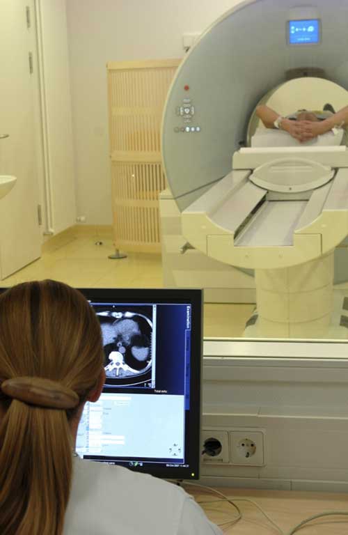
What does the equipment look like?
The CT scanner is typically a large, donut-shaped machine with a short tunnel in the center. You will lie on a narrow table that slides in and out of this short tunnel. Rotating around you, the x-ray tube and electronic x-ray detectors are located opposite each other in a ring, called a gantry. The computer workstation that processes the imaging information is in a separate control room. This is where the technologist operates the scanner and monitors your exam in direct visual contact. The technologist will be able to hear and talk to you using a speaker and microphone.
How does the procedure work?
In many ways, a CT scan works like other x-ray exams. Different body parts absorb x-rays in different amounts. This difference allows the doctor to distinguish body parts from one another on an x-ray or CT image.
A conventional x-ray exam directs a small amount of radiation through the body part under examination. A special electronic image recording plate captures the image. Bones appear white on the x-ray. Soft tissue, such as the heart or liver, shows up in shades of gray. Air appears black.
With CT scanning, several x-ray beams and electronic x-ray detectors rotate around you. These measure the amount of radiation being absorbed throughout your body. Sometimes, the exam table will move during the scan. A special computer program processes this large volume of data to create two-dimensional cross-sectional images of your body. The system displays the images on a computer monitor. CT imaging is sometimes compared to looking into a loaf of bread by cutting the loaf into thin slices. When the computer software reassembles the image slices, the result is a very detailed multidimensional view of the body's interior.
Nearly all CT scanners can obtain multiple slices in a single rotation. These multi-slice (multidetector) CT scanners obtain thinner slices in less time. This results in more detail.
Modern CT scanners can image large sections of the body in just a few seconds, and even faster in small children. Such speed is beneficial for all patients. Speed is especially beneficial for children, the elderly, and critically ill – anyone who finds it difficult to stay still, even for the brief time necessary to obtain images.
Some CT exams use a contrast material to enhance visibility in the body area under examination.
How is the procedure performed?
Prior to the procedure, you will be asked to drink several glasses of a liquid solution that contains a contrast material. The total amount of fluid you will need to drink is approximately 1 to 1.5 liters. You should inform your doctor if you think you will not be able to drink this amount of contrast. You will drink the contrast material over a period of approximately one hour in order to fill the long small intestine. The fluid expands the small bowel so that abnormalities can be seen better.
The technologist begins by positioning you on the CT exam table, usually lying flat on your back. They may use straps and pillows to help you maintain the correct position and remain still during the exam.
The exam may use contrast material, depending on the type of exam. If so, it will be swallowed, injected through an intravenous line (IV) or, rarely, administered by enema.
Next, the table will move quickly through the scanner to determine the correct starting position for the scans. Then, the table will move slowly through the machine for the actual CT scan. Depending on the type of CT scan, the machine may make several passes.
What will I experience during and after the procedure?
The technologist may ask you to hold your breath during the scanning. Any motion, including breathing and body movements, can lead to artifacts on the images. This loss of image quality can resemble the blurring seen on a photograph taken of a moving object.
When the exam is complete, the technologist will ask you to wait until they verify that the images are of high enough quality for accurate interpretation by the radiologist.
Though the scan is painless, you may have some discomfort from remaining still for several minutes or from placement of an IV. If you have a hard time staying still, are very nervous, anxious, or in pain, you may find a CT exam stressful. The technologist or nurse, under the direction of a doctor, may offer you some medication to help you tolerate the CT exam.
If the exam uses iodinated contrast material, your doctor will screen you for chronic or acute kidney disease. The doctor may administer contrast material intravenously (by vein), so you will feel a pin prick when the nurse inserts the needle into your vein. You may feel warm or flushed as the contrast is injected. You also may have a metallic taste in your mouth. This will pass. You may feel a need to urinate. However, these are only side effects of the contrast injection, and they subside quickly.
If you swallow oral contrast material, you may find the taste mildly unpleasant. However, most patients can easily tolerate it. If you receive an enema, you can expect to experience a sense of abdominal fullness. You may also feel an increasing need to expel the liquid. If so, be patient; the mild discomfort will not last long.
When you enter the CT scanner, you may see special light lines projected onto your body. These lines help ensure that you are in the correct position on the exam table. With modern CT scanners, you may hear slight buzzing, clicking and whirring sounds. These occur as the CT scanner's internal parts, not usually visible to you, revolve around you during the imaging process.
You will be alone in the exam room during the CT scan, unless there are special circumstances. For example, sometimes a parent wearing a lead shield may stay in the room with their child. However, the technologist will always be able to see, hear and speak with you through a built-in intercom system.
With pediatric patients, a parent may be allowed in the room but may need to wear a lead apron to minimize radiation exposure.
After a CT exam, the technologist will remove your intravenous line. They will cover the tiny hole made by the needle with a small dressing. You can return to your normal activities immediately.
The oral contrast material you will ingest for your enterography exam is not absorbed by the body and will be expelled through your stool. Therefore, loose stools will be present for a couple of hours after the examination. The oral contrast agent may cause nausea, diarrhea and abdominal cramps. You should tell your doctor if these mild side effects become severe or do not go away within a short time period.
See the Contrast Materials page for more information.
For children, the radiologist will adjust the CT scanner technique to their size and the area of interest to reduce the radiation dose.
Many scanners are fast enough to scan children without sedation. In special cases, children who cannot hold still may need sedation. Motion may cause blurring of the images and degrade image quality the same way that it affects photographs.
Who interprets the results and how do I get them?
A radiologist, a doctor specially trained to supervise and interpret radiology exams, will analyze the images. The radiologist will send an official report to the doctor who ordered the exam.
What are the benefits vs. risks?
Benefits
- CT scanning is painless, noninvasive, and accurate.
- A major advantage of CT is its ability to image bone, soft tissue, and blood vessels all at the same time.
- Unlike conventional x-rays, CT scanning provides very detailed images of many types of tissue as well as the lungs, bones, and blood vessels.
- CT exams are fast and simple. In emergency cases, they can reveal internal injuries and bleeding quickly enough to help save lives.
- CT has been shown to be a cost-effective imaging tool for a wide range of clinical problems.
- Compared to other imaging procedures of the small intestine, CT enterography is able to visualize the entire thickness of the bowel wall and to evaluate surrounding soft tissues. The other examinations, some of which are invasive, are only able to image the inner lining of the small intestine.
- CT enterography has been shown to diagnose and/or rule out certain conditions/diseases that could help determine your future medical care.
- CT enterography may eliminate the need for video capsule endoscopy (VCE) and the potential complications of that procedure.
- CT enterography allows other organs in the abdomen to be seen.
- CT is less sensitive to patient movement than MRI.
- Unlike MRI, an implanted medical device of any kind will not prevent you from having a CT scan.
- No radiation remains in a patient's body after a CT exam.
- The x-rays used for CT scanning should have no immediate side effects.
Risks
- There is always a slight chance of cancer from excessive exposure to radiation. However, the benefit of an accurate diagnosis far outweighs the risk involved with CT scanning.
- Women should always tell their doctor and x-ray or CT technologist if there is any chance they are pregnant. See the Safety in X-ray, Interventional Radiology and Nuclear Medicine Procedures page for more information about pregnancy and x-rays.
- Doctors do not generally recommend CT scanning for pregnant women unless medically necessary because of potential risk to the unborn baby.
- IV contrast manufacturers indicate mothers should not breastfeed their babies for 24-48 hours after contrast material is given. However, the most recent American College of Radiology (ACR) Manual on Contrast Media reports that studies show the amount of contrast absorbed by the infant during breastfeeding is extremely low. For further information please consult the ACR Manual on Contrast Media and its references.
- The risk of serious allergic reaction to contrast materials that contain iodine is extremely rare, and radiology departments are well-equipped to deal with them.
- Advancements in CT technology now allow CT enterography to be performed with even lower radiation doses.
- Because children are more sensitive to radiation, they should have a CT exam only if it is essential for making a diagnosis. They should not have repeated CT exams unless necessary. CT scans in children should always be done with low-dose technique.
What are the limitations of CT Enterography?
A person who is very large may not fit into the opening of a conventional CT scanner. Or, they may be over the weight limit—usually 450 pounds—for the moving table.
Certain bowel obstructions, small tumors and early inflammation may not be visualized with CT enterography. Another procedure, CT enteroclysis, provides greater filling and distension of the small intestine and may allow better detection of abnormalities. However, it requires placement of a tube into the small bowel through the nose.
This page was reviewed on June 01, 2022