Thyroid Scan and Uptake
Thyroid scan and uptake uses small amounts of radioactive materials called radiotracers, a special camera and a computer to provide information about your thyroid's size, shape, position and function that is often unattainable using other imaging procedures.
Tell your doctor if there's a possibility you are pregnant or if you are breastfeeding. Inform your doctor of any recent illnesses, medical conditions, allergies, medications you're taking and whether you've had any procedures within the last two months that used iodine-based contrast material. Your doctor will instruct you on how to prepare and may advise you not to eat for several hours prior to your exam. Leave jewelry at home and wear loose, comfortable clothing. You may be asked to wear a gown.
- What is a Thyroid Scan and Uptake?
- What are some common uses of the procedure?
- How should I prepare?
- What does the equipment look like?
- How does the procedure work?
- How is the procedure performed?
- What will I experience during and after the procedure?
- Who interprets the results and how do I get them?
- What are the benefits vs. risks?
- What are the limitations of the Thyroid Scan and Uptake?
What is a Thyroid Scan and Uptake?
A thyroid scan is a type of nuclear medicine imaging. The radioactive iodine uptake test (RAIU) is also known as a thyroid uptake. It is a measurement of thyroid function, but does not involve imaging.
Nuclear medicine uses small amounts of radioactive material called radiotracers. Doctors use nuclear medicine to diagnose, evaluate, and treat various diseases. These include cancer, heart disease, gastrointestinal, endocrine, or neurological disorders, and other conditions. Nuclear medicine exams pinpoint molecular activity. This gives them the potential to find disease in its earliest stages. They can also show whether you are responding to treatment.
Nuclear medicine is noninvasive. Except for intravenous injections, it is usually painless. These tests use radioactive materials called radiopharmaceuticals or radiotracers to help diagnose and assess medical conditions.
Radiotracers are molecules linked to, or "labeled" with, a small amount of radioactive material. They accumulate in tumors or regions of inflammation. They can also bind to specific proteins in the body. The most common radiotracer is F-18 fluorodeoxyglucose (FDG), a molecule similar to glucose. Cancer cells are more metabolically active and may absorb glucose at a higher rate. This higher rate can be seen on PET scans. This allows your doctor to detect disease before it may be seen on other imaging tests. FDG is just one of many radiotracers in use or in development.
You will usually receive the radiotracer in an injection. Or you may swallow it or inhale it as a gas, depending on the exam. It accumulates in the area under examination. A special camera detects gamma ray emissions from the radiotracer. The camera and a computer produce pictures and supply molecular information.
The thyroid scan and thyroid uptake provide information about the structure and function of the thyroid. The thyroid is a gland in the neck that controls metabolism, a chemical process that regulates the rate at which the body converts food to energy.
What are some common uses of the procedure?
The thyroid scan is used to determine the size, shape and position of the thyroid gland. The thyroid uptake is performed to evaluate the function of the gland. A whole-body thyroid scan is typically performed on people who have or had thyroid cancer.
A physician may perform these imaging tests to:
- determine if the gland is working properly
- help diagnose problems with the thyroid gland, such as an overactive thyroid gland, a condition called hyperthyroidism, cancer or other growths
- assess the nature of a nodule discovered in the gland
- detect areas of abnormality, such as lumps (nodules) or inflammation
- determine whether thyroid cancer has spread beyond the thyroid gland
- evaluate changes in the gland following medication use, surgery, radiotherapy or chemotherapy
How should I prepare?
You may wear a gown during the exam or be allowed to wear your own clothing.
Women should always tell their doctor and technologist if they are pregnant or breastfeeding. See the Radiation Safety page for more information about pregnancy and breastfeeding related to nuclear medicine imaging.
Tell the doctor and your exam technologist about any medications you are taking, including vitamins and herbal supplements. List any allergies, recent illnesses, and other medical conditions.
You should tell your physician if you:
- have had any tests, such as an x-ray or CT scan, surgeries or treatments using iodinated contrast material within the last two months.
- are taking medications or ingesting other substances that contain iodine, including kelp, seaweed, cough syrups, multivitamins or heart medications.
- have any allergies to iodine, medications and anesthetics.
- are breastfeeding.
In the days prior to your examination, blood tests may be performed to measure the level of thyroid hormones in your blood. You may be told not to eat for several hours before your exam because eating can affect the accuracy of the uptake measurement.
Leave jewelry and accessories at home or remove them prior to the exam. These objects may interfere with the procedure.
Your doctor will tell you how to prepare for your specific exam.
What does the equipment look like?
Nuclear medicine uses a special gamma camera and single-photon emission-computed tomography (SPECT) imaging techniques.
The gamma camera records the energy emissions from the radiotracer in your body and converts it into an image. The gamma camera itself does not emit any radiation. It has radiation detectors called gamma camera heads. These are encased in metal and plastic, often shaped like a box, and attached to a round, donut-shaped gantry. The patient lies on an exam table that slides in between two parallel gamma camera heads, above and beneath the patient. Sometimes, the doctor will orient the gamma camera heads at a 90-degree angle over the patient's body.
In SPECT, the gamma camera heads rotate around the patient's body to produce detailed, three-dimensional images.
A computer creates the images using the data from the gamma camera.
A probe is a small hand-held device resembling a microphone. It measures the amount of radiotracer in an area of your body.
How does the procedure work?
Ordinary x-ray exams pass x-rays through the body to create an image. Nuclear medicine uses radioactive materials called radiopharmaceuticals or radiotracers. Your doctor typically injects this material into your bloodstream. Or you may swallow it or inhale it as a gas. The material accumulates in the area under examination, where it gives off gamma rays. Special cameras detect this energy and, with the help of a computer, create pictures that detail how your organs and tissues look and function.
How is the procedure performed?
Doctors perform nuclear medicine exams on outpatients and hospitalized patients.
Thyroid Scan
You will lie on an exam table. If necessary, a nurse or technologist will insert an intravenous (IV) catheter into a vein in your hand or arm.
For most exams, you will receive an injection of the radiotracer. Or, you may swallow it or inhale it as a gas.
When radiotracer is taken by mouth, in either liquid or capsule form, it is typically swallowed up to 24 hours before the scan. The radiotracer given by intravenous injection is usually given up to 30 minutes prior to the test.
When it is time for the imaging to begin, you will lie down on a moveable examination table with your head tipped backward and neck extended. The gamma camera will then take a series of images, capturing images of the thyroid gland from three different angles. You will need to remain still for brief periods of time while the camera is taking pictures.
After the exam, you may need to wait until the technologist determines if more images are needed. Sometimes, the technologist takes more images to clarify or better visualize certain areas or structures. The need for more images does not necessarily mean there was a problem with the exam or that something is abnormal. It should not cause you concern.
If you have an intravenous (IV) line for the procedure, your technologist will usually remove it. The technologist will leave it in place if you are to have another procedure that same day that requires an IV line.
Actual scanning time for a thyroid scan is 30 minutes or less.
Thyroid Uptake
You will be given radioactive iodine (I-123 or I-131) in liquid or capsule form to swallow. The thyroid uptake will begin several hours to 24 hours later. Often, two separate uptake measurements are obtained at different times. For example, you may have uptake measurements at four to six hours and 24 hours.
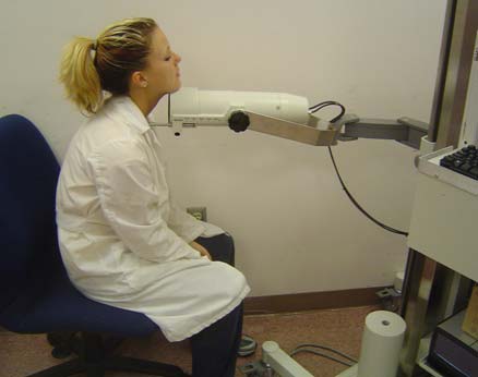
When it is time for the imaging to begin, you will sit in a chair facing a stationary probe positioned over the thyroid gland in the neck.
After the exam, you may need to wait until the technologist determines if more images are needed. Sometimes, the technologist takes more images to clarify or better visualize certain areas or structures. The need for more images does not necessarily mean there was a problem with the exam or that something is abnormal. It should not cause you concern.
Actual scanning time for each thyroid uptake is five minutes or less.
What will I experience during and after the procedure?
Most thyroid scan and thyroid uptake procedures are painless. However, during the thyroid scan, you may feel uncomfortable when lying completely still with your head extended backward while the gamma camera is taking images.
You will feel a slight pin prick when the technologist inserts the needle into your vein for the intravenous line. You may feel a cold sensation moving up your arm during the radiotracer injection. Generally, there are no other side effects.
Radiotracers have little or no taste. Inhaling a radiotracer feels no different than breathing the air around you or holding your breath.
It is important to remain still during the exam. Nuclear imaging causes no pain. However, having to remain still or in one position for long periods may cause discomfort.
Unless your doctor tells you otherwise, you may resume your normal activities after your exam. A technologist, nurse, or doctor will provide you with any necessary special instructions before you leave.
The small amount of radiotracer in your body will lose its radioactivity over time through the natural process of radioactive decay. It may also pass out of your body through your urine or stool during the first few hours or days after the test. Drink plenty of water to help flush the material out of your body.
Who interprets the results and how do I get them?
A radiologist or other doctor specially trained in nuclear medicine will interpret the images and send a report to your referring physician.
What are the benefits vs. risks?
Benefits
- Nuclear medicine exams provide unique information that is often unattainable using other imaging procedures. This information may include details on the function and anatomy of body structures.
- Nuclear medicine supplies the most useful diagnostic or treatment information for many diseases.
- A nuclear medicine scan is less expensive and may yield more precise information than exploratory surgery.
Risks
- Because nuclear medicine exams use only a small dose of radiotracer, they have a relatively low radiation exposure. This is acceptable for diagnostic exams. Thus, the potential benefits of an exam outweigh the very low radiation risk.
- Doctors have been using nuclear medicine diagnostic procedures for more than six decades. There are no known long-term adverse effects from such low-dose exposure.
- Your doctor always weighs the benefits of nuclear medicine treatment against any risks. Your doctor will discuss the significant risks prior to treatment and give you an opportunity to ask questions.
- Allergic reactions to radiotracers are extremely rare and usually mild. Always tell the nuclear medicine personnel about any allergies you may have. Describe any problems you may have had during previous nuclear medicine exams.
- The radiotracer injection may cause slight pain and redness. This should rapidly resolve.
- Women should always tell their doctor and radiology technologist if there is any possibility that they are pregnant, or they are breastfeeding. See the Radiation Safety page for more information about pregnancy, breastfeeding and nuclear medicine exams.
What are the limitations of the Thyroid Scan and Uptake?
The thyroid scan and thyroid uptake are not performed on patients who are pregnant because of the risk of exposing the fetus to radiation. These tests are also not recommended for breastfeeding women.
Nuclear medicine procedures can be time consuming. It can take several hours to days for the radiotracer to accumulate in the area of interest. Plus, imaging may take up to several hours to perform. In some cases, newer equipment can substantially shorten the procedure time.
The image resolution of nuclear medicine images may not be as high as that of CT or MRI. However, nuclear medicine scans are more sensitive for a variety of indications. The functional information they yield is often unobtainable using other imaging techniques.
Additional Information and Resources
RTAnswers.org: Radiation Therapy for Head and Neck Cancer
This page was reviewed on May 01, 2023