Cerebral Angiography
Cerebral angiography uses a catheter, x-ray imaging guidance and an injection of contrast material to examine blood vessels in the brain for abnormalities such as aneurysms and disease such as atherosclerosis (plaque). The use of a catheter makes it possible to combine diagnosis and treatment in a single procedure. Cerebral angiography produces very detailed, clear and accurate pictures of blood vessels in the brain and may eliminate the need for surgery.
Your doctor will instruct you on how to prepare, including any changes to your medication schedule. Tell your doctor if there's a possibility you are pregnant and discuss any recent illnesses, medical conditions, medications you're taking, and allergies, especially to iodinated contrast materials. If you're breastfeeding, ask your doctor how to proceed. If you are to be sedated, you may be told not to eat or drink anything for four to eight hours before your procedure. Also, you should plan to have someone drive you home. Leave jewelry at home and wear loose, comfortable clothing. You will be asked to wear a gown.
- What is Cerebral Angiography
- What are some common uses of the procedure?
- How should I prepare?
- What does the equipment look like?
- How does the procedure work?
- How is the procedure performed?
- What will I experience during and after the procedure?
- Who interprets the results and how do I get them?
- What are the benefits vs. risks?
- What are the limitations of Cerebral Angiography?
What is Cerebral Angiography
Angiography is a minimally invasive medical test that uses x-rays and an iodine-containing contrast material to produce pictures of blood vessels in the brain.
In cerebral angiography, a thin plastic tube called a catheter is inserted into an artery in the leg or arm through a small incision in the skin. Using x-ray guidance, the catheter is navigated to the area being examined. Once there, contrast material is injected through the tube and images are captured using ionizing radiation (x-rays).
Cerebral angiography is also called intra-arterial digital subtraction angiography (IADSA). This phrase refers to acquiring the images electronically, rather than with x-ray film. The images are electronically manipulated so that the overlying bone of the skull, normally obscuring the vessels, is removed from the image resulting in the remaining vessels being clearly seen.
What are some common uses of the procedure?
Physicians use the procedure to detect or confirm abnormalities within the blood vessels in the brain, including:
- an aneurysm, a bulge or sac that develops in an artery due to weakness of the arterial wall.
- atherosclerosis, a narrowing of the arteries.
- arteriovenous malformation, a tangle of dilated blood vessels that disrupts normal blood flow in the brain.
- vasculitis, an inflammation of the blood vessels, generally narrowing them.
- a brain tumor.
- a blood clot.
- a tear in the wall of an artery, known as a vascular dissection.
- a stroke.
A cerebral angiogram may be performed:
- to evaluate arteries of the head and neck before surgery.
- to provide additional information on abnormalities seen on MRI or CT of the head, such as the blood supply to a tumor.
- to prepare for other medical treatment, such as in the surgical removal of a tumor.
- in preparation for minimally invasive treatment of a vessel abnormality.
The procedure may also be used to help diagnose the cause of symptoms, such as:
- severe headaches
- slurred speech
- dizziness
- blurred or double vision
- weakness or numbness
- loss of coordination or balance.
How should I prepare?
Tell your doctor about all the medications you take, including herbal supplements. List any allergies, especially to local anesthetic, general anesthesia, or contrast materials. Your doctor may tell you to stop taking aspirin, nonsteroidal anti-inflammatory drugs (NSAIDs) or blood thinners before your procedure.
Tell your doctor about recent illnesses or other medical conditions.
If you are to receive a sedative during the procedure, the doctor may tell you not to eat or drink anything for four to eight hours before your exam. If you are sedated, have someone accompany you and drive you home afterward.
For more information about sedation, visit the Anesthesia page.
You will receive specific instructions on how to prepare, including any changes you need to make to your regular medication schedule.
Your doctor will likely tell you not to eat or drink anything after midnight before your procedure. Your doctor will tell you which medications you may take in the morning.
Women should always tell their doctor and technologist if they are pregnant. Doctors will not perform many tests during pregnancy to avoid exposing the fetus to radiation. If an x-ray is necessary, the doctor will take precautions to minimize radiation exposure to the baby. See the Radiation Safety page for more information about pregnancy and x-rays.
If you are breastfeeding at the time of the exam, you should ask your radiologist how to proceed. It may help to pump breast milk ahead of time and keep it on hand for use after contrast material has cleared from your body, about 24 hours after the test.
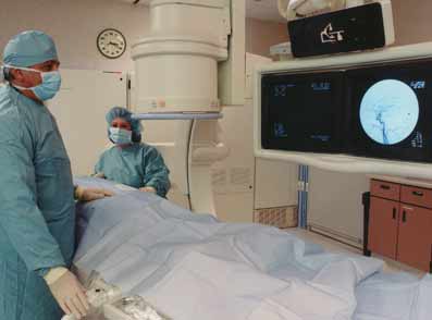
What does the equipment look like?
In this procedure, x-ray equipment will be used.
A catheter is a long, thin plastic tube that is considerably smaller than a "pencil lead." It is about 1/8 inch in diameter.
A catheter is inserted by a radiologist, usually through a tiny hole made by a needle in an artery in the groin. Using x-ray guidance, it is painlessly guided through the body to various vessels in the neck that supply blood to the brain.
This procedure may use other equipment, including an intravenous line (IV), ultrasound machine and devices that monitor your heart beat and blood pressure.
How does the procedure work?
X-rays are a form of radiation like light or radio waves. X-rays pass through most objects, including the body. The technologist carefully aims the x-ray beam at the area of interest. The machine produces a small burst of radiation that passes through your body. The radiation records an image on photographic film or a special detector.
Different parts of the body absorb the x-rays in varying degrees. Dense bone absorbs much of the radiation while soft tissue (muscle, fat, and organs) allow more of the x-rays to pass through them. As a result, bones appear white on the x-ray, soft tissue shows up in shades of gray, and air appears black.
Most x-ray images are electronically stored digital files. Your doctor can easily access these stored images to diagnose and manage your condition.
Fluoroscopy uses a continuous or pulsed x-ray beam to create images and project them onto a video monitor. Your exam may use a contrast material to clearly define the area of interest. Fluoroscopy allows your doctor to view joints or internal organs in motion. The exam also captures still images or movies and stores them electronically on a computer.
How is the procedure performed?
This procedure is often done on an outpatient basis. However, some patients may require admission following the procedure. Ask your doctor if you will need to be admitted.
Prior to your procedure, your doctor may test your blood to check your kidney function and to determine if your blood clots normally.
Because the cerebral angiogram and recovery period may last for several hours, you will be asked to empty your bladder before the procedure begins.
A nurse or technologist will insert an intravenous (IV) line into a vein in your hand or arm to administer a sedative. This procedure may use moderate sedation. It does not require a breathing tube. However, some patients may require general anesthesia.
In children up to mid-teens, cerebral angiography is usually performed with the patient under general anesthesia.
The doctor or nurse will attach devices to your body to monitor your heart rate and blood pressure.
You will lie on the procedure table.
Your head will be held in place using a strap, tape or a foam head holder so you cannot move it during the procedure.
The nurse will sterilize the area of your body where the catheter is to be inserted. They will sterilize and cover this area with a surgical drape.
Your doctor will numb the area with a local anesthetic. This may briefly burn or sting before the area becomes numb.
The doctor will make a very small skin incision at the site.
Using x-ray-guidance, a catheter (a long, thin, hollow plastic tube) is inserted into a blood vessel through a tiny hole made by a needle and directed to the area to be examined.
The contrast material is then injected through the catheter. A special machine, called a power injector, is used to deliver the contrast material at a precise rate and volume. The injector is attached to the catheter for this purpose. When the contrast material reaches the blood vessels being examined, several sets of x-rays will be taken.
When the procedure is complete, the doctor will remove the catheter and apply pressure to stop any bleeding. Sometimes, your doctor may use a closure device to seal the small hole in the artery. This will allow you to move around more quickly. No stitches are visible on the skin. The nurse will cover this tiny opening in the skin with a dressing.
The doctor or nurse will remove your IV line before you go home.
The procedure is usually completed within one to three hours. Additional time may be required for exam preparation, setup and post-procedure care.
What will I experience during and after the procedure?
You will feel a slight pinch when the nurse inserts the needle into your vein for the IV line and when they inject the local anesthetic. Most of the sensation is at the skin incision site. The doctor will numb this area using local anesthetic. You may feel pressure when the doctor inserts the catheter into the vein or artery. However, you will not feel serious discomfort.
If the procedure uses sedation, you will feel relaxed, sleepy, and comfortable. You may or may not remain awake, depending on how deeply you are sedated.
You may feel slight pressure when the doctor inserts the catheter, but no serious discomfort.
As the contrast material passes through your body, you may feel warm. This will quickly pass.
You will be asked to remain very still while the x-ray images are taken.
The most difficult part of the procedure may be lying flat for several hours.
Once the procedure is complete, the catheter will be removed by the radiologist. To prevent any bleeding from the puncture site, it must be closed either by placing pressure to the site or by applying a vascular closure device which plugs the puncture site directly. Pressure may be either applied by hand or with a special clamp which takes about 10 minutes for the tiny hole in the artery to close. If the radiologist determines that a vascular closure device can be placed, a small plug is inserted as the catheter is removed and quickly seals the puncture site, decreasing the time required for you to lie flat after the procedure.
You will remain in the recovery room for observation for a few hours following the procedure before you return home.
If the catheter was placed in the groin area, you will be given specific instructions regarding how long you may need to keep your leg straight. This will vary based on the technique used to repair the hole created in order to insert the catheter. You may apply ice to the site where the catheter was inserted to relieve pain and swelling.
You may resume your normal diet immediately after the exam. You will be able to resume all other normal activities eight to 12 hours after the exam.
You should report to your physician immediately if you experience any of the following after your procedure:
- weakness or numbness in the muscles of your face, arms or legs
- slurred speech
- vision problems
- signs of infection at the catheter site
- dizziness
- chest pain
- difficulty breathing
- rash
- difficulty in using the extremity where the puncture/incision was made
Who interprets the results and how do I get them?
A radiologist, a doctor specifically trained to perform, supervise, and interpret radiology examinations, will analyze the images. They will send a signed report to your primary care or referring physician, who will share the results with you.
You may need a follow-up exam. If so, your doctor will explain why. Sometimes a follow-up exam further evaluates a potential issue with more views or a special imaging technique. It may also see if there has been any change in an issue over time. Follow-up exams are often the best way to see if treatment is working or if a problem needs attention.
What are the benefits vs. risks?
Benefits
- Angiography may eliminate the need for surgery. If surgery remains necessary, it can be performed more accurately.
- Cerebral angiography presents a very detailed, clear and accurate picture of blood vessels in the brain. This is especially helpful when a surgical procedure or other treatment is being considered.
- Results from cerebral angiography are more accurate than those produced by carotid Doppler ultrasound or other noninvasive imaging of the blood vessels.
- Use of a catheter makes it possible to combine diagnosis and treatment in a single procedure.
- No radiation stays in your body after an x-ray exam.
- X-rays usually have no side effects in the typical diagnostic range for this exam.
Risks
- There is always a slight chance of cancer from excessive exposure to radiation. However, given the small amount of radiation used in medical imaging, the benefit of an accurate diagnosis far outweighs the associated risk.
- There is a very slight risk of an allergic reaction if the procedure uses an injection of contrast material.
- If you have a history of allergy to x-ray contrast material, your radiologist may advise that you take special medication for 24 hours before cerebral angiography to lessen the risk of allergic reaction. However, the risk of an allergic reaction from contrast material injected into an artery is less than if it is introduced into a vein.
- Women should always tell their doctor and x-ray technologist if they are pregnant. See the Radiation Safety page for more information about pregnancy and x-rays.
- Nursing mothers should wait for 24 hours after contrast material injection before resuming breastfeeding.
- The risk of serious allergic reaction to contrast materials that contain iodine is extremely rare, and radiology departments are well equipped to deal with them.
- If you have diabetes or kidney disease, the kidneys may be injured due to the contrast material. In most cases, the kidneys will regain their normal function within five to seven days.
- Any procedure that places a catheter inside a blood vessel carries certain risks. These risks include damage to the blood vessel, bruising or bleeding at the puncture site, and infection. The doctor will take precautions to mitigate these risks.
- There is a small risk that blood will form a clot around the tip of the catheter, blocking the artery and making it necessary to operate to reopen the vessel.
- There is a risk of stroke with this procedure if the catheter dislodges plaque from a vessel wall that blocks blood flow within the brain. Although stroke may be a complication associated with cerebral angiography, it is uncommon.
- Rarely, the catheter punctures the artery, causing internal bleeding. It also is possible that the catheter tip will separate material from the inner lining of the artery, causing a block downstream in the blood vessel. Given that children do not usually have plaque in their arteries, they would not be as susceptible as adults to have such a complication.
A Word About Minimizing Radiation Exposure
Doctors take special care during x-ray exams to use the lowest radiation dose possible while producing the best images for evaluation. National and international radiology protection organizations continually review and update the technique standards radiology professionals use.
Modern x-ray systems minimize stray (scatter) radiation by using controlled x-ray beams and dose control methods. This ensures that the areas of your body not being imaged receive minimal radiation exposure.
When performing cerebral angiography in children or young adults, care is often taken to minimize radiation to the ovaries and testes by placing a lead drape under the pelvis.
What are the limitations of Cerebral Angiography?
Patients with impaired kidney function may not be good candidates for this procedure.
Patients who have previously had allergic reactions to iodine-containing x-ray contrast materials are at risk of having a second reaction to similar contrast agents.
This page was reviewed on November 01, 2022