General Biopsy
A biopsy is the removal of tissue from any part of the body to examine it for disease. Some may remove a small tissue sample with a needle while others may surgically remove a suspicious nodule or lump.
Most needle biopsies are performed on an outpatient basis with minimal preparation. Your doctor will give you instructions based on the type of biopsy being performed. Tell your doctor if there's a possibility you are pregnant. Discuss any medications you're taking, including blood thinners such as aspirin and herbal supplements, and whether you have any allergies – especially to anesthesia. Your physician will advise you to stop taking blood thinners for a specific period of time before your procedure, and you may be told not to eat or drink anything for eight hours beforehand. Leave jewelry at home and wear loose, comfortable clothing. You may be asked to wear a gown.
- What are biopsies?
- What are some common uses of the procedure?
- How should I prepare?
- What does the equipment look like?
- How does the procedure work?
- How is the procedure performed?
- What will I experience during and after the procedure?
- Who interprets the results and how do I get them?
- What are the benefits vs. risks?
- What are the limitations of biopsies?
What are biopsies?
A biopsy is the removal of tissue to examine it for disease. The tissue samples can be taken from any part of the body. Biopsies are performed in several different ways. Some biopsies involve removing a small amount of tissue with a needle while others involve surgically removing an entire lump, or nodule, that is suspicious.
Often, the tissue is removed by placing a needle through the skin (percutaneously) to the area of abnormality. Biopsies can be safely performed with imaging guidance such as ultrasound, computed tomography (CT), or magnetic resonance imaging (MRI). These types of imaging are used to determine exactly where to place the needle and perform the biopsy.
What are some common uses of the procedure?
When your doctor finds a nodule, they may order imaging tests to help determine if it is benign (non-cancerous) or malignant (cancerous). If imaging exams cannot clearly define the abnormality, a biopsy may be necessary.
Usually, a biopsy is performed to examine tissue for disease. Biopsies are frequently used to diagnose cancer, but they can help identify other conditions such as infections and inflammatory and autoimmune disorders. They may also be done to match organ tissue before a transplant and to look for signs of organ rejection following a transplant.
Biopsies are performed in many areas of the body and for many reasons. The following is a list of common biopsy types and why they may be necessary:
- Abdominal biopsy is used to diagnose whether a lump in the abdomen is cancerous or benign. The lumps can be located in the fat deep within the abdomen or in abdominal organs. A sample of the lump is removed percutaneously under image guidance (ultrasound or CT), or surgically using a laparoscope or by open surgery.
- Bone biopsy is used to diagnose cancer or infection in the bones. This type of biopsy can be performed through the skin (percutaneously) with a needle or surgically.
- Bone marrow biopsy is used to diagnose cancer in the blood, such as leukemia. A small sample of the bone and bone marrow are removed using a needle. Sometimes, only the bone marrow is removed for examination.
- Breast biopsy is used to determine if a lump in the breast is cancerous or benign. It can be performed a number of ways:
- Stereotactic (mammographically-guided)
- Ultrasound-guided
- MRI-guided
- Endometrial biopsy may be used when looking for the cause of abnormal uterine bleeding, to examine the lining of the uterus and to diagnose cancer. This type of biopsy can be performed by using a small needle-like device to capture a sample or by using a tool to scrape some of the lining for examination.
- Kidney (renal) biopsy is used to examine the condition of a kidney with kidney failure, inflammation in the kidney or a suspected tumor (such as cancer). It can also be used to examine a transplanted kidney for signs of transplant rejection. Kidney biopsies are performed with image guidance (ultrasound or CT) using a needle to remove a small sample of the tissue.
- Liver biopsy is used to diagnose diseases of the liver such as hepatitis C, cirrhosis, infections and cancer. It can also be used to examine a transplanted liver for signs of transplant rejection. This is a frequent indication in children. Liver biopsies are typically performed percutaneously by inserting a needle through the skin. The liver can also be biopsied via a catheter inserted through the jugular vein (a large neck vein) to capture a tissue sample, or can be biopsied surgically.
- Lung or chest nodule biopsy is performed when an abnormality of the lung is visible on an x-ray or CT scan. Lung biopsies can be performed through bronchoscopy by inserting an instrument called a bronchoscope through the patient's mouth and into the airway to reach the area to be biopsied, through the skin by inserting a needle percutaneously, or by surgically removing the lump.
- Lymph node biopsy is performed whenever there are enlarged or abnormal lymph nodes. They can be performed with a needle or surgically.
- Muscle biopsy is used to diagnose infections that affect muscle, defects in the muscle and diseases of the connective tissue and blood vessels. This type of biopsy can be performed using a needle or surgically.
- Nerve biopsy is used to examine damage to small nerves, degeneration and destruction of the nerve and inflammatory nerve conditions. Nerve biopsies are typically performed surgically.
- Skin biopsy examines a growth or an area on the skin, such as a mole, that has changed its appearance. Skin biopsies can be performed by shaving a small sample of the skin, removing a sample with a scalpel or by way of an instrument used to punch through a portion of the skin.
- Testicular biopsy is used to determine underlying causes of male infertility. It is also used to determine if a lump in the testicles is cancerous or benign. Testicular biopsies can be performed using a needle, by a small cut made in the skin or surgically. Testicular needle biopsy is rarely used to diagnose testicular cancer since it increases the likelihood that cancer will spread. An ultrasound is usually used to diagnose testicular cancer, and the affected testicle is often removed through open surgery called inguinal orchiectomy.
- Thyroid biopsy is used to find the cause of a nodule in the thyroid gland. This type of biopsy is typically performed using a needle with ultrasound guidance.
Almost any organ can be biopsied, including the bladder, heart, neck, prostate, parathyroid glands, etc.
How should I prepare?
Most needle biopsies are performed in an outpatient setting with minimal preparation. When you schedule your biopsy appointment, you will receive detailed instructions about preparation for the biopsy procedure.
If you are having a needle or surgical biopsy, ask your health care provider if you need to stop taking any medications before the procedure.
Your doctor may tell you not to eat or drink for eight hours before your biopsy. However, you may take your routine medications with sips of water. If you are diabetic and take insulin, ask your doctor if you need to adjust your usual insulin dose.
Prior to a needle biopsy, tell your doctor about all the medications you take, including herbal supplements. List any allergies, especially to anesthesia. Your doctor may tell you to stop taking aspirin or a blood thinner for a time before your procedure.
Also, tell your doctor about recent illnesses and other medical conditions.
You may need to change into a gown for the procedure.
Women should always tell their doctor if there is any possibility that they are pregnant. Doctors do not perform some procedures that use image-guidance during pregnancy because radiation can be harmful to the fetus. See the Radiation Safety page for more information about pregnancy and x-rays.
You may want to have someone accompany you and drive you home afterward. This will be necessary if you receive sedation.
Preparation for a biopsy procedure will be similar for children. If your child is undergoing a biopsy procedure, the physician will provide you with instructions.
What does the equipment look like?
There are many different types of biopsy procedures. The equipment used for each type of biopsy will vary depending on the type of procedure.
In needle biopsy, a sample of tissue or fluid is removed with a needle and sent to the laboratory for further analysis.
A biopsy needle is generally several inches long. The barrel is about as wide as a large paper clip. The needle is hollow so it can capture the tissue specimen.
A biopsy may use one of several types of needles. Common ones include:
- A fine needle attached to a syringe, smaller than needles typically used to draw blood.
- A core needle, also called an automatic, spring-loaded needle, which consists of an inner needle connected to a trough, or shallow receptacle, covered by a sheath and attached to a spring-loaded mechanism.
- A vacuum-assisted device (VAD), which uses a vacuum to aid in obtaining larger pieces of tissue.
Doctors perform needle biopsies with the guidance of computed tomography (CT), fluoroscopy, ultrasound, or MRI.
A mammography unit is a box with a tube that produces x-rays. The unit is used exclusively for breast x-ray exams and features special accessories to limit x-ray exposure to only the breast. The unit features a device to hold and compress the breast and position it so the technologist can capture images at different angles.
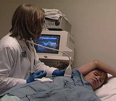
Ultrasound machines consist of a computer console, video monitor and an attached transducer. The transducer is a small hand-held device that resembles a microphone. Some exams may use different transducers (with different capabilities) during a single exam. The transducer sends out inaudible, high-frequency sound waves into the body and listens for the returning echoes. The same principles apply to sonar used by boats and submarines.
The technologist applies a small amount of gel to the area under examination and places the transducer there. The gel allows sound waves to travel back and forth between the transducer and the area under examination. The ultrasound image is immediately visible on a video monitor. The computer creates the image based on the loudness (amplitude), pitch (frequency), and time it takes for the ultrasound signal to return to the transducer. It also considers what type of body structure and/or tissue the sound is traveling through.
The CT scanner is typically a large, donut-shaped machine with a short tunnel in the center. You will lie on a narrow table that slides in and out of this short tunnel. Rotating around you, the x-ray tube and electronic x-ray detectors are located opposite each other in a ring, called a gantry. The computer workstation that processes the imaging information is in a separate control room. This is where the technologist operates the scanner and monitors your exam in direct visual contact. The technologist will be able to hear and talk to you using a speaker and microphone.
A vacuum-assisted device (VAD) is a vacuum-powered instrument that uses pressure to pull tissue into the needle.
In some situations, image guidance is used to help a surgeon to perform a surgical biopsy. A thin guide wire may be placed or marking dye injected with x-ray, ultrasound, or CT guidance to aid the surgeon in locating the correct area for a surgical biopsy.
Other sterile equipment that may be involved in a biopsy procedure includes syringes, sponges, forceps, scalpels and a specimen cup or microscope slide.
How does the procedure work?
The type of procedure used to perform the biopsy depends on the location of the tissue that needs to be examined.
Most areas of the body can be biopsied with a needle device. This is the least invasive option, and usually allows for the patient to return home the same day. Imaging guidance with x-ray, ultrasound, CT or MRI allows for accurate placement of the needle to locate the best place to take a tissue sample.
In hard to reach places, biopsies using surgery in a hospital operating room may sometimes be necessary. A surgeon will perform surgery to remove the tissue needed for the biopsy. The surgeon may use an instrument with a camera to help locate the best place to biopsy and remove the tissue sample.
Using imaging guidance, the doctor inserts the needle through the skin and advances it into the lesion.
They will remove tissue samples using one of several methods.
- In a fine needle aspiration, a fine gauge needle and a syringe withdraw fluid or clusters of cells.
- In a core needle biopsy, the automated mechanism moves the needle forward and fills the needle trough, or shallow receptacle, with “cores” of tissue. The outer sheath instantly moves forward to cut the tissue and keep it in the trough. This process is repeated several times.
- In a vacuum-assisted biopsy, the doctor inserts the needle into the site of abnormality. They activate the vacuum device, which pulls the tissue into the needle trough, cuts it with the sheath, and retracts it through the hollow core of the needle. The doctor may repeat this procedure several times.
How is the procedure performed?
Imaging-guided, minimally invasive procedures, such as needle biopsies, are most often performed by a specially trained radiologist, an interventional radiologist or a neuroradiologist.
Doctors usually perform needle biopsies on an outpatient basis.
A nurse or technologist may insert an intravenous (IV) line into a vein in your hand or arm. This will allow them to provide sedation or relaxation medication intravenously during the procedure. You may also receive a mild sedative prior to the biopsy.
The doctor will use a local anesthetic to numb the path of the needle.
Some biopsies, such as breast or thyroid biopsies, may be performed without sedation. The nurse or technologist will advise you at the time of the procedure regarding the sedation.
When a biopsy is performed on a child, it is more likely that general anesthesia will be required to keep them comfortable during the procedure.
If the doctor is using fluoroscopy guidance, you will lie down or stand for the procedure.
If the doctor is using CT or MRI guidance, you will lie down during the procedure. They will use a limited CT or MRI scan to confirm the location of the nodule and the safest approach for the targeted area. Once they confirm the nodule’s location, they will mark the entry site on the skin. The doctor will clean and disinfect the skin around the insertion site and cover it with a clean and sterile drape.
The doctor will make a very small nick in the skin at the site where the biopsy needle will be inserted.
Using imaging guidance, the doctor will insert the needle through the skin, advance it to the site of the nodule, and remove samples of tissue. They may need to collect several specimens for complete analysis.
After the sampling, the doctor will remove the needle.
Once the biopsy is complete, the doctor will apply pressure to stop any bleeding and cover the opening in the skin with a dressing. No sutures are needed.
You may be taken to an observation area for several hours. The doctor may use X-ray(s) or other imaging tests to monitor for complications.
Biopsy procedures are typically performed the same way for children.
For stereotactic breast biopsies, you may lie face down or sit up on a moveable exam table and the affected breast or breasts will be positioned into openings in the table.
In a fine needle aspiration, a fine gauge needle and a syringe withdraw fluid or clusters of cells.
In a core needle biopsy, the automated mechanism is activated, moving the needle forward and filling the needle trough, or shallow receptacle, with 'cores' of breast tissue. The outer sheath instantly moves forward to cut the tissue and keep it in the trough. This process may be repeated several times.
In some breast biopsies, the tissue is removed with a vacuum-assisted device (VAD). Vacuum pressure is used to pull tissue from the breast through the needle into the sampling chamber. Without withdrawing and reinserting the needle, it rotates positions and collects additional samples. Typically, several samples of tissue are collected from around the lesion. After this sampling, the needle will be removed.
If a surgical biopsy is being performed, a wire may be inserted into the suspicious area as a guide for the surgeon.
A small marker may be placed at the site, so that it can be located in the future if necessary.
Once the biopsy is complete, pressure will be applied to stop any bleeding, and the skin will be covered with a dressing or bandage.
Depending on the type of biopsy performed, you may be able to return home immediately after the procedure.
This procedure is usually completed within one hour. You may be required to stay in an area for observation for several hours after the biopsy depending on the type of biopsy performed.
What will I experience during and after the procedure?
When you receive the local anesthetic to numb the skin, you will feel a slight pin prick from the needle. The area will become numb within a short time.
After the skin is numb, you may feel some pressure when the biopsy needle is inserted and when the biopsy sample is being taken.
You may receive a mild sedative prior to the biopsy. If needed, you may receive sedation or relaxation medication intravenously during the procedure.
You may feel sore at the area of the biopsy for a few days. Your doctor can prescribe pain relief medication if you have significant pain from the biopsy.
Aftercare instructions vary. However, you generally may remove your bandage one day after the procedure, and you may bathe or shower as normal.
Who interprets the results and how do I get them?
After the tissue is collected, it is sent to a laboratory for analysis. A pathologist will examine the biopsy tissue under a microscope. A full report from the pathologist will be sent to your doctor in a few days.
You should ask the doctor performing the procedure how you will receive your results.
If you underwent a breast biopsy, it is likely the radiologist will discuss your results with you.
Your interventional radiologist may recommend a follow-up visit.
This visit may include a physical check-up, imaging exam(s), and blood tests. During your follow-up visit, tell your doctor if you have noticed any side effects or changes.
What are the benefits vs. risks?
- Needle biopsy is a reliable way to obtain tissue samples that can help diagnose whether a nodule is benign (non-cancerous) or malignant.
- A needle biopsy is less invasive than open and closed surgical biopsies, both of which involve a larger incision in the skin and local or general anesthesia.
- Generally, the procedure is not painful. Results are as accurate as when a tissue sample is removed surgically.
- Recovery time is brief, and patients can soon resume their usual activities.
- Any procedure that penetrates the skin carries a risk of infection. The chance of infection requiring antibiotic treatment appears to be less than one in 1,000.
Generally, a biopsy procedure is safe and causes minimal injury. Complications that may result from biopsies include:
- Bleeding
- Infection
- Accidental injury to adjacent structures such as the bowel during abdominal biopsy or lung parenchyma during renal biopsy
What are the limitations of biopsies?
In some cases, the amount of tissue obtained from a needle biopsy may not be sufficient and the biopsy may have to be repeated. This may be particularly true with trying to make a diagnosis of lymphoma.
Rarely, less invasive breast biopsy procedures may be unable to detect some lesions or determine the extent of disease present. If the diagnosis remains uncertain after a technically successful procedure, surgical biopsy will usually be necessary.
Any imaging-guided procedure will not be able to be used unless the area of abnormality can be seen. Some lesions, such as clustered calcifications on mammography are not as clearly shown with ultrasound as they are with mammography. Therefore, stereotactic biopsy is usually used in breast imaging to biopsy calcifications. Fluoroscopy sometimes will not be able to locate chest nodules, and CT will be used for guidance. Your radiologist will use the image guidance best suited to biopsy the area in question.
This page was reviewed on April 01, 2024