Breast Lumps
A breast lump is a mass that develops in the breast. Breast lumps vary in size and texture and may cause pain. Some are not found until a physical or imaging exam. Most breast lumps are benign (non-cancerous).
Your doctor will likely perform a physical exam to evaluate a breast lump. To determine whether that lump is benign, your doctor will likely order a mammogram and breast ultrasound. In addition, breast MRI, PET/CT or scintimammography may be obtained. If the lump is confirmed to be benign, no further action may be needed, but your doctor may want to monitor it to see if it changes, grows or disappears over time. If the tests are inconclusive, a biopsy using ultrasound, x-ray or magnetic resonance imaging guidance may be performed. If the lump is confirmed to be cancer, surgery is usually performed. Additional treatment may include radiation therapy, chemotherapy, or hormone therapy.
What are breast lumps?
A breast lump is a mass that develops in the breast. Depending on the type, breast lumps may be large or small and may feel hard or spongy. Some lumps cause pain, while others go unnoticed until identified during an imaging test.
A lump may be discovered by a woman doing breast self-exam or by her health care provider during a physical exam. Suspicious lumps may also be detected during annual screening mammography. Although uncommon, breast lumps can occur in men.
It is important to become familiar with how your breasts normally look and feel so that you are able to report any changes to your doctor.
How are breast lumps diagnosed and evaluated?
Most breast lumps are benign (not cancer). Proving that a lump is not cancer often involves imaging tests. One or more of the following imaging tests may be performed:
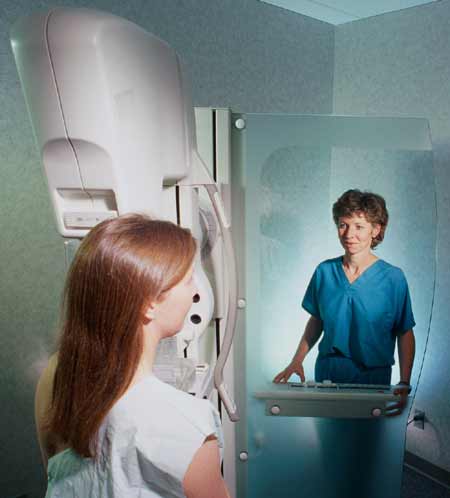
- mammogram: Mammography uses low dose x-rays to examine the breasts. This type of imaging involves exposing the breasts to a small amount of ionizing radiation to obtain pictures of the inside of the breasts. Either two single images or two tomosynthesis images (also called 3-D mammography) are taken of each breast to begin the evaluation. Additional images may be needed. See the Safety page for more information about x-rays.
- breast ultrasound: Breast ultrasound uses sound waves to create pictures of the inside of the breasts. Breast ultrasound can capture images of areas of the breast that may be difficult to see with mammography. It can also help to determine whether a breast lump is solid or fluid.
- breast MRI: Breast MRI uses a powerful magnetic field, radiofrequency pulses and a computer to produce detailed pictures of the inside of the breasts. MRI is helpful in evaluating breast lumps that are not visible with mammography or ultrasound—although it may not be appropriate for all women. Your doctor will help determine if breast MRI is right for you. Breast MRI requires injection of contrast material.
If a lump is proven to be benign by its appearance on these exams, no further steps may need to be taken. Your doctor may want to monitor the area at future visits to check if the breast lump has changed, grown or gone away.
If these tests do not clearly show that the lump is benign, a biopsy may be necessary. One of the following image-guided procedures may be performed:
- ultrasound-guided biopsy: During this type of biopsy, using ultrasound imaging to find the lump, a radiologist will administer local anesthesia, and then advance a thin sampling needle into the lump to remove some tissue for evaluation under a microscope. The biopsy procedure is usually quick, but it may take a few days before the final tissue analysis (pathology report) is ready.
- stereotactic (x-ray guided) biopsy: During this type of biopsy, using a digital mammography x-ray machine to image the area of concern, a radiologist will administer local anesthesia and then position a sampling needle at this site to remove thin tissue samples for further evaluation.
- MRI-guided biopsy: During this type of biopsy, using an MRI machine to localize the area of concern, a radiologist will administer local anesthesia and then position a sampling needle at this site to remove thin tissue samples for further evaluation.
Often, the radiologist will place a tiny metal marker (approximately the size of one sesame seed) in the area where tissue was sampled so that any residual lump will not need additional testing if it is seen on future mammograms.
If you need a biopsy, it is important to choose a facility with expertise, preferably one where the radiologists specialize in breast imaging. One measure of a facility's expertise in breast biopsy can be found in its ACR accreditation status. Check the facilities in your area by searching the ACR-accredited facilities database.
How are breast lumps treated?
If a lump is proven to be cancer, surgery is usually performed. The surgeon will explain appropriate surgical options and provide you with the information necessary to make this decision.
You may have several consultations with other physicians for additional treatment, including radiation therapy and chemotherapy or hormone therapy.
One of the following radiation therapy treatments may be used after surgery to ensure any microscopic cancer cells are eliminated:
- External Beam Therapy
- Intensity-Modulated Radiation Therapy (IMRT)
- Brachytherapy (Interstitial Therapy)
For more information visit the Breast Cancer disease and Breast Cancer Treatment pages.
This page was reviewed on April 15, 2022