Pediatric Voiding Urosonography
Pediatric contrast-enhanced voiding urosonography uses ultrasound to examine a child's bladder and urinary tract. It is often used after a urinary tract infection to check if urine is backing up into the kidneys.
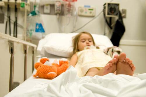
Little to no special preparation is required and sedation is rarely needed. Explain what will happen during the exam to your child. This will help ease anxiety and any confusion about what is expected.
- What is Pediatric Contrast-enhanced Voiding Urosonography?
- What are some common uses of the procedure?
- How should we prepare for the procedure?
- What does the equipment look like?
- How does the procedure work?
- How is the procedure performed?
- What will my child experience during and after the procedure?
- Who interprets the results and how do we get them?
- What are the benefits vs. risks?
- What are the limitations of the procedure?
What is Pediatric Contrast-enhanced Voiding Urosonography?
Pediatric contrast-enhanced voiding urosonography (ceVUS) examines the bladder and urinary tract with ultrasound. During the exam, liquid contrast material is introduced into the bladder through a catheter. The liquid helps show what may be causing pain or problems with urination. Ultrasound is particularly suitable for children because it does not use radiation.
What are some common uses of the procedure?
Doctors often use ceVUS after a urinary tract infection to check for vesicoureteral reflux (VUR). Kidneys produce urine which flows through tubes called ureters to the bladder. As the bladder fills, a valve prevents urine from backing up into the kidneys. During urination, urine leaves the bladder through the urethra.
Some children have a valve or ureter problem that allows urine to flow backwards. This is called VUR. In mild cases, urine backs up into the lower ureter. In severe cases, it can back up into the kidney. Children with VUR are usually born with it. Other causes include:
- blockage in the bladder
- abnormal urination with very high pressure within the bladder
- incomplete emptying of the bladder
The only symptom of VUR may be a urinary tract infection.
Pediatric ceVUS may be used to detect blockages in the urinary tract caused by valves or focal narrowing. It also helps assess problems such as incomplete emptying of the bladder.
How should we prepare for the procedure?
Tell your doctor if your child has recently been sick. Discuss any medical conditions, medications, and allergies, especially to contrast material. Your child will have to remove their clothing and wear a gown. Little to no special preparation is required. Sedation is rarely needed. Tell your child what will happen during the exam so there will be no confusion about what is expected.
What does the equipment look like?
Ultrasound machines consist of a computer console, video monitor and an attached transducer. The transducer is a small hand-held device that resembles a microphone. Some exams may use different transducers (with different capabilities) during a single exam. The transducer sends out inaudible, high-frequency sound waves into the body and listens for the returning echoes. The same principles apply to sonar used by boats and submarines.
The technologist applies a small amount of gel to the area under examination and places the transducer there. The gel allows sound waves to travel back and forth between the transducer and the area under examination. The ultrasound image is immediately visible on a video monitor. The computer creates the image based on the loudness (amplitude), pitch (frequency), and time it takes for the ultrasound signal to return to the transducer. It also considers what type of body structure and/or tissue the sound is traveling through.
The bladder is filled with contrast material using a flexible, hollow plastic tube called a catheter. The catheter has a diameter smaller than the urethra.
How does the procedure work?
Ultrasound imaging uses the same principles as the sonar that bats, ships, and fishermen use. When a sound wave strikes an object, it bounces back or echoes. By measuring these echo waves, it is possible to determine how far away the object is as well as its size, shape, and consistency. This includes whether the object is solid or filled with fluid.
Doctors use ultrasound to detect changes in the appearance of organs, tissues, and vessels and to detect abnormal masses, such as tumors.
In an ultrasound exam, a transducer both sends the sound waves and records the echoing (returning) waves. When the transducer is pressed against the skin, it sends small pulses of inaudible, high-frequency sound waves into the body. As the sound waves bounce off internal organs, fluids and tissues, the sensitive receiver in the transducer records tiny changes in the sound's pitch and direction. A computer instantly measures these signature waves and displays them as real-time pictures on a monitor. The technologist typically captures one or more frames of the moving pictures as still images. They may also save short video loops of the images.
Microbubble Contrast Materials
The contrast material used in ceVUS is made up of tiny bubbles of gas held in a supporting shell. The bubbles are smaller than a red blood cell. They appear brighter on ultrasound because they reflect ultrasound waves. Ultrasound technology captures differences between the bubbles in the bladder and urinary tract and the surrounding tissues. These differences produce an image with increased contrast. This allows doctors to track the flow of urine and more easily identify any problems.
How is the procedure performed?
This exam is usually done on an outpatient basis.
The technologist begins by placing the child on the table. A parent can be next to the child and help them to lie still during the imaging.
For most ultrasound exams, your child will be positioned lying face-up on an exam table. Patients may be turned to either side to improve the quality of the images. They may also be asked to lie face-down.
The technologist applies a clear water-based gel to the body area under examination. This helps the transducer make secure contact with the body. It also helps eliminate air pockets between the transducer and the skin that can block the sound waves from passing into your body. The technologist or radiologist places the transducer on the skin in various locations, sweeping over the area of interest. They may also angle the sound beam from a different location to better see an area of concern.
The bladder and kidneys are scanned with ultrasound. After the genital area is cleaned, the catheter is inserted through the urethra. The catheter may be taped to the skin so that it will not come loose during the procedure. The bladder is then filled with a liquid mixture of normal saline and contrast material. When the bladder is full, the child will urinate while lying on the table. The liquid will be collected with a urinal, bed pan or absorbent pad. The child may also be allowed to sit on a potty while being scanned from the back.
The radiologist or technologist will use ultrasound to monitor the bladder during filling and urination. They will check to see if any of the liquid contrast material goes backward into one or both ureters and kidneys. They will also check the bladder and urethra to see if their shape, lumen, and contour are normal.
After the exam, the radiologist or technologist may ask you and your child to wait until they confirm that all the necessary images have been obtained.
What will my child experience during and after the procedure?
Placing a tube in the bladder can be uncomfortable. The antiseptic used to clean and prepare for the catheter insertion may feel cold. Most children accept the procedure after it is completely explained.
Parents may be allowed to stay in the exam room to comfort their child.
The transducer does not usually cause discomfort when it is pressed against the area being examined. However, if the area being scanned is tender, the transducer may cause your child to feel pressure or minor pain.
Once the imaging is complete, the clear ultrasound gel will be wiped off your child's skin. The ultrasound gel does not usually stain or discolor clothing.
Although the time may vary, the entire procedure will generally take less than 30 minutes.
After the exam, your child should be able to resume their normal activities immediately.
Who interprets the results and how do we get them?
A radiologist will analyze the images and send a signed report to your pediatrician or referring physician. That doctor will discuss the results with you.
A follow-up exam may be needed. Your doctor will explain the exact reason why. Sometimes another exam is done to further examine a problem. Or, it may be used to see if there has been a change. Follow-up exams are sometimes the best way to see if treatment is working.
What are the benefits vs. risks?
Benefits
- Ultrasound imaging is extremely safe and does not use any radiation.
- Ultrasound scanning gives a clear picture of many organs that do not show up well on a regular x-ray.
- Pediatric ceVUS provides your doctor with valuable, detailed information to assess for the presence or absence of vesicoureteral reflux which can be seen in children with a urinary tract infection.
- Pediatric ceVUS helps your doctor decide if treatment is needed. Some conditions do not require treatment. Other conditions may require medication. Some may even need surgery.
Risks
- Some children may have discomfort urinating after the procedure. This usually goes away in less than 12 hours.
What are the limitations of the procedure?
ceVUS is not recommended for children who have an active, untreated urinary tract infection.
Also, ceVUS cannot be performed in children whose kidneys are not well visualized on ultrasound imaging due to body habitus or size.
This page was reviewed on November 01, 2022