Abdominal Contrast Enhanced Ultrasound (CEUS)
Abdominal contrast-enhanced ultrasound (CEUS) uses gas-filled microbubbles to better visualize organs and blood vessels within the abdomen and pelvis. This exam may evaluate the liver, spleen, kidneys, pancreas, bowel, and/or bladder.
This procedure requires little to no special preparation. Your doctor will tell you how to prepare, including whether you should not eat or drink beforehand. Wear loose, comfortable clothing. You may need to wear a gown.
- What is Abdominal Contrast Enhanced Ultrasound (CEUS)?
- What are some common uses of the procedure?
- How should I prepare?
- What does the equipment look like?
- How does the procedure work?
- How is the procedure performed?
- What will I experience during and after the procedure?
- Who interprets the results and how do I get them?
- What are the benefits vs. risks?
- What are the limitations of Abdominal Contrast Enhanced Ultrasound?
What is Abdominal Contrast Enhanced Ultrasound (CEUS)?
CEUS combines ultrasound of the abdomen with a special type of intravenous contrast agent to improve the visualization of blood vessels and organs. CEUS helps the sonographer and the radiologist see abnormalities in the organs or blood vessels. Doctors use this same contrast agent to better assess the heart.
Ultrasound imaging is a noninvasive medical test that helps physicians diagnose and treat medical conditions. It is safe and painless. It produces pictures of the inside of the body using sound waves. Ultrasound imaging is also called sonography. It uses a small probe called a transducer and gel placed directly on the skin. High-frequency sound waves travel from the probe through the gel into the body. The probe collects the sounds that bounce back. A computer uses those sound waves to create an image. Ultrasound exams do not use radiation (x-rays). Because ultrasound captures images in real-time, it can show the structure and movement of the body's internal organs. The images can also show blood flowing through blood vessels.
Abdominal CEUS uses a small amount of contrast material made up of gas-filled microbubbles. The doctor injects this material into the bloodstream through an intravenous (IV) line. Since these tiny microbubbles are similar in size to red blood cells, they pass through the heart and lungs and into arterial circulation. The microbubbles travel via the blood to the organs. Contrast material improves the visualization of organs (and any masses or tumors they may contain) by highlighting blood vessels. This is similar but slightly different than other contrast materials used in CT scans and MRI. Abdominal CEUS is particularly useful for patients who may have kidney failure. This is because the microbubbles can be safely given in all patients, including those with severe kidney disease or those patients on dialysis. Microbubble contrast agents for CEUS are also safe to use in patients who are allergic to the contrast materials used in CT and MRI. CEUS allows you to safely receive more than one contrast material injection during the same exam. This lets your sonographer and radiologist examine multiple lesions or multiple organs during the same session if necessary.
What are some common uses of the procedure?
Doctors use abdominal CEUS to evaluate the:
Doctors also use abdominal CEUS to help diagnose a wide variety of conditions, such as:
- liver lesions discovered during routine ultrasound
- liver lesions that are not well seen on CT or MRI scans
- blood flow abnormalities in the liver
- cirrhosis of the liver
- kidney lesions
- traumatic injuries to the abdomen
- lesions of the spleen
How should I prepare?
Wear comfortable, loose-fitting clothing. You may need to remove all clothing and jewelry in the area to be examined.
You may need to change into a gown for the procedure.
Preparations will depend on the type of ultrasound exam they are going to do.
- For most exams, you do not need any kind of preparation
- For an exam of the pancreas, you may be asked to fast up to 4 hours before
- For a bowel exam, it may be best to avoid carbonated drinks up to 4 hours before
What does the equipment look like?
Ultrasound machines consist of a computer console, video monitor and an attached transducer. The transducer is a small hand-held device that resembles a microphone. Some exams may use different transducers (with different capabilities) during a single exam. The transducer sends out inaudible, high-frequency sound waves into the body and listens for the returning echoes. The same principles apply to sonar used by boats and submarines.
The technologist applies a small amount of gel to the area under examination and places the transducer there. The gel allows sound waves to travel back and forth between the transducer and the area under examination. The ultrasound image is immediately visible on a video monitor. The computer creates the image based on the loudness (amplitude), pitch (frequency), and time it takes for the ultrasound signal to return to the transducer. It also considers what type of body structure and/or tissue the sound is traveling through.
How does the procedure work?
Ultrasound imaging uses the same principles as the sonar that bats, ships, and fishermen use. When a sound wave strikes an object, it bounces back or echoes. By measuring these echo waves, it is possible to determine how far away the object is as well as its size, shape, and consistency. This includes whether the object is solid or filled with fluid.
Doctors use ultrasound to detect changes in the appearance of organs, tissues, and vessels and to detect abnormal masses, such as tumors.
In an ultrasound exam, a transducer both sends the sound waves and records the echoing (returning) waves. When the transducer is pressed against the skin, it sends small pulses of inaudible, high-frequency sound waves into the body. As the sound waves bounce off internal organs, fluids and tissues, the sensitive receiver in the transducer records tiny changes in the sound's pitch and direction. A computer instantly measures these signature waves and displays them as real-time pictures on a monitor. The technologist typically captures one or more frames of the moving pictures as still images. They may also save short video loops of the images.
Doppler ultrasound, a special ultrasound technique, measures the direction and speed of blood cells as they move through vessels. The movement of blood cells causes a change in pitch of the reflected sound waves (called the Doppler effect). A computer collects and processes the sounds and creates graphs or color pictures that represent the flow of blood through the blood vessels.
Doctors often use CEUS instead of non-contrast ultrasound because it can show the difference between normal and diseased tissues. In the case of cancers, this difference is primarily due to cancers having more blood flow. Cancers may appear brighter than normal tissue or clear the blood faster than surrounding normal tissue on CEUS. However, image interpretation requires expertise, as noncancerous conditions may also have a similar contrast pattern.
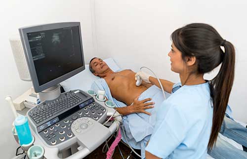
How is the procedure performed?
For most ultrasound exams, you will lie face-up on an exam table that can be tilted or moved. You may need to turn to either side or lie face-down to improve the quality of the images.
The technologist applies a clear water-based gel to the body area under examination. This helps the transducer make secure contact with the body. It also helps eliminate air pockets between the transducer and the skin that can block the sound waves from passing into your body. The technologist or radiologist places the transducer on the skin in various locations, sweeping over the area of interest. They may also angle the sound beam from a different location to better see an area of concern.
Once the initial ultrasound scan is complete, you will receive a small injection of contrast material through an intravenous (IV) catheter placed in the arm or hand. An injection of saline will push the contrast agent out of the IV catheter and into the body.
You will lie still for several minutes while the sonographer or radiologist uses the transducer to capture images of the bubbles traveling through the organ and/or blood vessel of interest. Occasionally, the doctor will repeat the injection.
After about 10 minutes, all the bubbles will have popped harmlessly. The small amount of gas they contain leaves the body through normal breathing.
What will I experience during and after the procedure?
You may feel some pain when the doctor inserts the intravenous catheter into your hand or arm. Some patients experience a cold sensation or slight discomfort when the saline is injected. Problems with the microbubble contrast itself are extremely rare, but allergic reactions have been reported. Occasionally, patients may experience a mild temporary headache or unusual taste in their mouth.
There is usually no discomfort from pressure as the transducer is pressed against the area being examined. However, if the sonographer scans an area of tenderness, you may feel pressure or minor pain from the transducer.
Once the imaging is complete, the sonographer will wipe the clear ultrasound gel off your skin. The ultrasound gel does not usually stain or discolor clothing.
The entire CEUS procedure will generally take 30-60 minutes. This time may vary depending on the number of lesions your doctor evaluates.
When the exam is complete, the technologist may ask you to dress and wait while they review the ultrasound images.
After an ultrasound exam, you should be able to resume your normal activities immediately.
Who interprets the results and how do I get them?
A radiologist, a physician specifically trained to supervise and interpret medical imaging exams, will analyze the images. They will also send a signed report to the doctor who requested the exam. Sometimes, the radiologist will discuss the results with you after the exam. CEUS frequently requires that the radiologist to review the images again while writing their report. This helps them arrive at a definitive diagnosis. In this case, you can get the results from the doctor who ordered the exam once they receive the signed report.
Follow-up exams may be necessary. Your doctor will explain the exact reason why another exam is necessary. Sometimes a follow-up exam is done because a potential abnormality needs further evaluation with additional views or another radiology test. A follow-up exam may also be necessary to monitor a known abnormality over time for any change. Follow-up exams are sometimes the best way to see if a treatment is working or if an abnormality is changing over time.
What are the benefits vs. risks?
Benefits
- An ultrasound exam may occasionally be uncomfortable, but it is rarely painful.
- Ultrasound is widely available, easy-to-use, and less expensive than other imaging methods.
- Ultrasound imaging is extremely safe and does not use any radiation.
- Ultrasound scanning gives a clear picture of many organs that do not show up well on a regular x-ray.
- Abdominal CEUS may eliminate the need for other tests such as CT and/or MRI. This can be especially helpful for patients who have claustrophobia or a hard time laying still.
Risks
- The contrast agents used in abdominal CEUS carry a very small risk of an allergic reaction. This risk is about the same as the risk of allergic reaction to many antibiotics. In fact, it is lower than the risk of allergic reaction to CT and MRI contrast materials.
What are the limitations of Abdominal Contrast Enhanced Ultrasound?
Air can disrupt ultrasound waves. Therefore, a bowel full of air may limit the effectiveness of an ultrasound scan. Ultrasound does not image inside our lungs well for the same reason.
Large or obese patients are more difficult to image by ultrasound. This is because greater amounts of tissue weaken the sound waves as they pass into the body.
Usually only one region of the body can be examined per injection, and only a few areas can be examined in one exam. Because of this, CEUS is not always suitable for looking at multiple organs at once.
Certain lesions and areas of interest may be difficult to image, including lesions less than 1 centimeter, and lesions that are close to the lung or deep within the liver.
This page was reviewed on November 01, 2022