Breast Tomosynthesis
Breast tomosynthesis is an advanced form of mammography, a specific type of breast imaging that uses low-dose x-rays to detect cancer early when it is most treatable. Breast tomosynthesis is not yet available in all imaging facilities.
Tell your doctor about any breast symptoms or problems, prior surgeries, hormone use, whether you have a family or personal history of breast cancer, and if there's a possibility you are pregnant. If possible, obtain copies of your prior mammograms and make them available to your radiologist on the day of your exam. Leave jewelry at home and wear loose, comfortable clothing. You may be asked to wear a gown. Don't wear deodorant, talcum powder or lotion under your arms or on your breasts as these may appear on the mammogram and interfere with correct diagnosis.
- What is breast tomosynthesis?
- What are some common uses of the procedure?
- How should I prepare?
- What does the equipment look like?
- How does the procedure work?
- How is the procedure performed?
- What will I experience during and after the procedure?
- Who interprets the results and how do I get them?
- What are the benefits vs. risks?
- What are the limitations of breast tomosynthesis?
What is breast tomosynthesis?
Breast tomosynthesis, also called three-dimensional (3-D) mammography and digital breast tomosynthesis (DBT), is an advanced form of breast imaging, or mammography, that uses a low-dose x-ray system and computer reconstructions to create three-dimensional images of the breasts. Breast tomosynthesis aids in the early detection and diagnosis of breast disease.
An x-ray exam helps doctors diagnose and treat medical conditions. It exposes you to a small dose of ionizing radiation to produce pictures of the inside of the body. X-rays are the oldest and most often used form of medical imaging.
While mammography is the best screening tool for breast cancer available today, it does not detect all breast cancers. Breast tomosynthesis overcomes some of the limitations of standard mammography, but it is not yet available in all imaging facilities.
A conventional x-ray examination of the breast, called a mammogram, is two-dimensional: two x-ray images are taken of the breast, from top-to-bottom and from angled side-to-side, while the breast is compressed between a clear plastic paddle and an imaging detector. Although compression is necessary to obtain breast images, it may cause overlapping of the breast tissue in which abnormal tissue can be hidden and superimposed normal tissue can appear abnormal.
In breast tomosynthesis, the x-ray tube moves in an arc over the compressed breast capturing multiple images of each breast from different angles. These digital images are then reconstructed or "synthesized" into a set of three-dimensional images by a computer. These three dimensional image sets help minimize the tissue overlap that can hide cancers or make it difficult to distinguish normal overlapping breast tissue from tumors.
What are some common uses of the procedure?
Breast tomosynthesis can be used to perform screening mammography, a test to detect early breast cancer in women experiencing no symptoms. Screening mammography with breast tomosynthesis has been shown to improve accuracy and reduce false positive rates in women of all breast densities. The largest improvements with tomosynthesis are seen in women with dense breast tissue, but women with non-dense breasts also demonstrate significantly better cancer detection and significantly fewer false positive findings when tomosynthesis is used. See the Dense Breasts page for more information about dense breasts.
Breast tomosynthesis may also be used to perform diagnostic mammography to detect and diagnose breast disease in women experiencing symptoms such as a lump, pain, skin dimpling or nipple discharge.
Screening Mammography with Breast Tomosynthesis
Mammography plays a central part in the early detection of breast cancers because it can often show changes in the breast years before a patient or physician can feel them. Current guidelines from the American College of Radiology (ACR) recommend screening mammography every year for women beginning at age 40. Research has shown that annual mammograms lead to early detection of breast cancers when they are most curable and breast-conservation therapies are available. See the Mammography and Breast Cancer Screening pages for more information.
Diagnostic Mammography with Breast Tomosynthesis
Diagnostic mammography is used to evaluate a patient with abnormal clinical findings—such as a breast lump or nipple discharge—that have been found by a patient or physician. Diagnostic mammography may also be done after an abnormal screening mammogram in order to evaluate the area of concern on the screening exam.
How should I prepare?
Before scheduling a mammogram, the American Cancer Society (ACS) and other specialty organizations recommend that you discuss any new findings or problems in your breasts with your doctor. In addition, inform your doctor of any prior surgeries, hormone use, and family or personal history of breast cancer.
Do not schedule your screening mammogram for the week before your menstrual period if your breasts are usually tender during this time. The best time for a screening mammogram is one week following your period. Always inform your doctor or x-ray technologist if there is any possibility that you are pregnant.
The ACS also recommends you:
- Do not wear deodorant, talcum powder or lotion under your arms or on your breasts on the day of the exam. These can be confused for calcium deposits on the mammogram.
- Describe any breast symptoms or problems to the technologist performing the exam.
- Obtain your prior mammograms and make them available to the radiologist if they were done at a different location. This is needed for comparison with your current exam and can often be obtained on a CD.
- Ask when your results will be available; do not assume the results are normal if you do not hear from your doctor or the mammography facility.
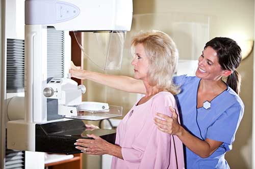
What does the equipment look like?
A mammography unit is a box with a tube that produces x-rays. The unit is used exclusively for breast x-ray exams and features special accessories to limit x-ray exposure to only the breast. The unit features a device to hold and compress the breast and position it so the technologist can capture images at different angles.
Breast tomosynthesis is performed using a digital mammography unit, but not all digital mammography machines are equipped to perform tomosynthesis imaging.
How does the procedure work?
Breast tomosynthesis uses a low-dose x-ray system, electronics and a computer to convert x-ray images of the breast to a three-dimensional image set. Multiple x-ray images of the breast are digitized using systems similar to those found in digital cameras and then transferred to a computer where they are reconstructed or "synthesized" into a three-dimensional image set. In this way, 3-D breast imaging is similar to computed tomography (CT) imaging in which a series of thin high-resolution "slices" are assembled together to create a 3-D reconstruction of the body.
X-rays are a form of radiation like light or radio waves. X-rays pass through most objects, including the body. The technologist carefully aims the x-ray beam at the area of interest. The machine produces a small burst of radiation that passes through your body. The radiation records an image on photographic film or a special detector.
Different parts of the body absorb the x-rays in varying degrees. Dense bone absorbs much of the radiation while soft tissue (muscle, fat, and organs) allow more of the x-rays to pass through them. As a result, bones appear white on the x-ray, soft tissue shows up in shades of gray, and air appears black.
Most x-ray images are electronically stored digital files. Your doctor can easily access these stored images to diagnose and manage your condition.
How is the procedure performed?
Breast tomosynthesis is performed on an outpatient basis.
During this exam, a specially qualified radiologic technologist will position your breast in the mammography unit. Your breast will be placed on a special platform and gradually compressed with a clear plastic paddle. Breast compression is necessary during tomosynthesis imaging in order to:
- Even out the breast thickness so that all of the tissue can be visualized.
- Spread out the tissue so that small abnormalities are less likely to be hidden by overlying breast tissue.
- Allow the use of a lower x-ray dose since a thinner amount of breast tissue is being imaged.
- Hold the breast still in order to minimize blurring of the image caused by motion.
Your breast will remain compressed while the x-ray tube moves from one side of your breast to the other in an arc, capturing multiple images from different angles along the way. In addition to the three-dimensional image set, the breast tomosynthesis exam can also synthesize standard two-dimensional images of the breast for interpretation.
You must hold very still and may need to hold your breath for a few seconds while the technologist takes the x-ray. This helps reduce the possibility of a blurred image. The technologist will walk behind a wall or into the next room to activate the x-ray machine.
When the examination is complete, the technologist may ask you to wait until the radiologist confirms they have all the necessary images.
The examination process should take about 30 minutes.
What will I experience during and after the procedure?
You will feel pressure on your breast as it is squeezed by the compression paddle. Some women with sensitive breasts may experience discomfort. If this is the case, schedule the procedure when your breasts are least tender. Be sure to inform the technologist if pain occurs as compression is increased. If discomfort is significant, less compression will be used. Always remember compression allows better quality mammograms, including breast tomosynthesis exams.
Who interprets the results and how do I get them?
A radiologist, a doctor trained to supervise and interpret radiology examinations, will analyze the images. The radiologist will send a signed report to your primary care or referring physician who will discuss the results with you.
You will also be notified of the results by the mammography facility.
You may need a follow-up exam. If so, your doctor will explain why. Sometimes a follow-up exam further evaluates a potential issue with more views or a special imaging technique. It may also see if there has been any change in an issue over time. Follow-up exams are often the best way to see if treatment is working or if a problem needs attention.
What are the benefits vs. risks?
Benefits
- Imaging of the breast allows detection of small tumors. When cancers are small, the woman has more treatment options.
- The use of screening mammography increases the detection of small abnormal tissue growths confined to the milk ducts in the breast, called ductal carcinoma in situ (DCIS). These early tumors rarely harm patients if they are removed at this stage and mammography is an excellent way to detect these tumors. It is also useful for detecting all types of breast cancer, including invasive ductal and invasive lobular cancer.
- No radiation stays in your body after an x-ray exam.
- X-rays usually have no side effects in the typical diagnostic range for this exam.
- Large population studies have shown that screening with breast tomosynthesis results in improved breast cancer detection rates and fewer "call-backs," instances where women are called back from screening for additional testing because of a potentially abnormal finding.
- Breast tomosynthesis may also result in:
- earlier detection of small breast cancers that may be hidden on a conventional mammogram
- greater accuracy in pinpointing the size, shape and location of breast abnormalities
- fewer unnecessary biopsies or additional tests
- greater likelihood of detecting multiple breast tumors
- clearer images of abnormalities within dense breast tissue
Risks
- There is always a slight chance of cancer from excessive exposure to radiation. However, given the small amount of radiation used in medical imaging, the benefit of an accurate diagnosis far outweighs the associated risk.
- The radiation dose for this procedure varies. See the Radiation Dose page for more information.
- False Positive Mammograms. Five percent to 15 percent of screening mammograms require more testing such as additional mammograms or ultrasound. Most of these tests turn out to be normal. If there is an abnormal finding, a follow-up or biopsy may have to be performed. Most biopsies confirm that no cancer is present. The likelihood of false positive findings and negative biopsies is reduced when tomosynthesis imaging is used for screening and diagnostic mammographic examinations.
- Women should always tell their doctor and x-ray technologist if they are pregnant. See the Radiation Safety page for more information about pregnancy and x-rays.
A Word About Minimizing Radiation Exposure
Doctors take special care during x-ray exams to use the lowest radiation dose possible while producing the best images for evaluation. National and international radiology protection organizations continually review and update the technique standards radiology professionals use.
Modern x-ray systems minimize stray (scatter) radiation by using controlled x-ray beams and dose control methods. This ensures that the areas of your body not being imaged receive minimal radiation exposure.
Although the radiation dose for breast tomosynthesis is slightly higher than the dosage used in standard mammography, it remains within the FDA-approved safe levels for radiation from mammograms. Some systems have doses very similar to conventional mammography.
What are the limitations of breast tomosynthesis?
Interpretations of mammograms can be difficult because a normal breast looks different for each woman. Also, the appearance of an image may be challenging if you have undergone breast surgery. Because some breast cancers are hard to visualize, a radiologist may want to compare the image to views from previous examinations. It is very important to realize that not all breast cancers can be seen on mammography.
While mammography is the best screening tool for breast cancer available today, mammograms do not detect all breast cancers. This is called a false negative result. On the other hand, when a mammogram looks abnormal and no cancer is present, this is called a false-positive result.
Advanced imaging techniques such as breast tomosynthesis are helping to overcome the limitations of conventional mammography. Research continues on breast tomosynthesis and other breast imaging techniques that can contribute to the early detection of breast cancer and improve the accuracy in distinguishing non-cancerous breast conditions from breast cancers.
Breast implants may also impede accurate mammogram readings because both silicone and saline implants are not transparent on x-rays and can block a clear view of the tissues behind them, especially if the implant has been placed in front of the chest muscles. Experienced technologists and radiologists know how to carefully compress the breasts and move the implants out of the image to improve the view as much as possible without rupturing the implant.
Additional Information and Resources
Mammography Saves Lives: Breast Imaging
ACR: Breast Density and Breast Cancer Screening Brochure
MedLinePlus: Mammography
This page was reviewed on March 11, 2024