

Image/Video Gallery
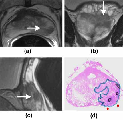
T2-weighted MR images with arrows pointing to low signal intensity area i.e. suspicious for cancer in the prostate gland. The representative sections show the area suspicious for cancer in the prostate gland at different planes i.e. axial (a), coronal (b), and sagittal (c). The histopathology slice (d), with cancer outlined, closely corresponds to the MR image.
Note: Images are shown for illustrative purposes. Do not attempt to draw conclusions or make diagnoses by comparing these images to other medical images, particularly your own. Only qualified physicians should interpret images; the radiologist is the physician expert trained in medical imaging.