

Image/Video Gallery
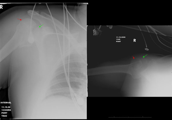
Frontal and axillary views of the right shoulder demonstrate the humeral head (red arrow) to be displaced out of the glenoid socket (green arrow) and migrated posteriorly compatible with a posterior shoulder dislocation. This patient had bilateral posterior shoulder dislocation from electrocution (very rare case, the other shoulder is not shown here).
Note: Images are shown for illustrative purposes. Do not attempt to draw conclusions or make diagnoses by comparing these images to other medical images, particularly your own. Only qualified physicians should interpret images; the radiologist is the physician expert trained in medical imaging.