

Image/Video Gallery
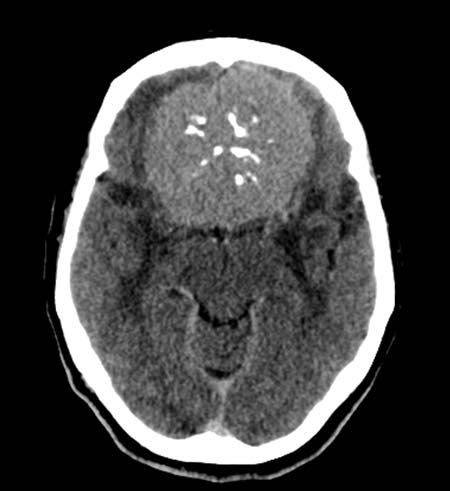
Meningioma. Non-contrast head CT scan shows a large, midline frontal mass, which is slightly hyperdense relative to brain, and has a number of small, relatively central calcifications, but only a mild degree of surrounding edema (swelling). The minimal edema suggests a slowly growing neoplasm is present.
Note: Images are shown for illustrative purposes. Do not attempt to draw conclusions or make diagnoses by comparing these images to other medical images, particularly your own. Only qualified physicians should interpret images; the radiologist is the physician expert trained in medical imaging.