Image/Video Gallery

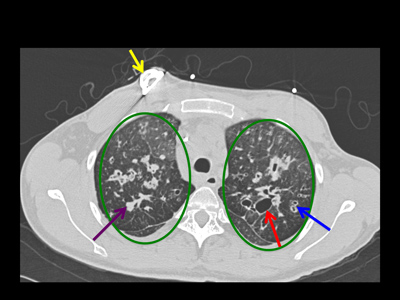
Axial computed tomography (CT) of the upper chest showing a port catheter (yellow arrow) in place to administer chronic IV medicine. Dilated airways (red arrows) are seen as black areas within the lungs (green ovals), some with thickened walls (blue arrows). Mucous (purple arrow), which is white in appearance, is also seen in some of the airways.
Note: Images are shown for illustrative purposes. Do not attempt to draw conclusions or make diagnoses by comparing these images to other medical images, particularly your own. Only qualified physicians should interpret images; the radiologist is the physician expert trained in medical imaging.


