

Image/Video Gallery
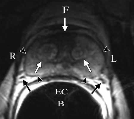
Endorectal MRI of the anatomy of the prostate using a clinical MR scanner. The anterior fibromuscular stroma (arrow) consists of nonglandular tissue and appears dark. The muscular stroma layer in the posterior prostate base is denoted by arrowheads.
F = Front
R = Right
L = Left
B = Back
EC = Endorectal Coil
Note: Images are shown for illustrative purposes. Do not attempt to draw conclusions or make diagnoses by comparing these images to other medical images, particularly your own. Only qualified physicians should interpret images; the radiologist is the physician expert trained in medical imaging.