Last reviewed on March 19, 2025
Musculoskeletal Ultrasound
Ultrasound imaging uses sound waves to produce pictures of muscles, tendons, ligaments, nerves and joints throughout the body. It is used to help diagnose sprains, strains, tears, trapped nerves, arthritis and other musculoskeletal conditions. Ultrasound is safe, noninvasive, and does not use ionizing radiation.
This procedure requires little to no special preparation. Leave jewelry at home and wear loose, comfortable clothing. You may be asked to wear a gown.
What is Ultrasound Imaging of the Musculoskeletal System?
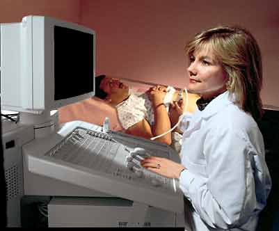
Ultrasound imaging is a noninvasive medical test that helps physicians diagnose and treat medical conditions. It is safe and painless. It produces pictures of the inside of the body using sound waves. Ultrasound imaging is also called sonography. It uses a small probe called a transducer and gel placed directly on the skin. High-frequency sound waves travel from the probe through the gel into the body. The probe collects the sounds that bounce back. A computer uses those sound waves to create an image. Ultrasound exams do not use radiation (x-rays). Because ultrasound captures images in real-time, it can show the structure and movement of the body's internal organs. The images can also show blood flowing through blood vessels.
Ultrasound images of the musculoskeletal system provide pictures of muscles, tendons, ligaments, joints, nerves and soft tissues throughout the body.
What are some common uses of the procedure?
Ultrasound images are typically used to help diagnose:
- tendon tears or tendinitis of the rotator cuff in the shoulder, Achilles tendon in the ankle and many other tendons throughout the body.
- muscle tears, masses or fluid collections.
- ligament sprains or tears.
- inflammation or fluid (effusions) within the bursae and joints.
- early changes of rheumatoid arthritis.
- nerve entrapments such as carpal tunnel syndrome.
- benign and malignant soft tissue tumors.
- ganglion cysts.
- hernias.
- foreign bodies in the soft tissues (such as splinters or glass).
- dislocations and dysplasia of the hip in infants.
- fluid in a painful hip joint in children.
- neck muscle abnormalities in infants with torticollis (neck twisting).
- soft tissue masses (lumps/bumps) in children.
Who interprets the results and how do I get them?
A radiologist, a doctor trained to supervise and interpret radiology exams, will analyze the images. The radiologist will send a signed report to the doctor who requested the exam. Your doctor will then share the results with you. In some cases, the radiologist may discuss results with you after the exam.
You may need a follow-up exam. If so, your doctor will explain why. Sometimes a follow-up exam further evaluates a potential issue with more views or a special imaging technique. It may also see if there has been any change in an issue over time. Follow-up exams are often the best way to see if treatment is working or if a problem needs attention.
What are the benefits vs. risks?
Benefits
- Most ultrasound scanning is noninvasive (no needles or injections).
- Occasionally, an ultrasound exam may be temporarily uncomfortable, but it should not be painful.
- Ultrasound is widely available, easy to use, and less expensive than most other imaging methods.
- Ultrasound imaging is extremely safe and does not use radiation.
- Ultrasound scanning gives a clear picture of soft tissues that do not show up well on x-ray images.
- Ultrasound provides real-time imaging. This makes it a good tool for guiding minimally invasive procedures such as needle biopsies and fluid aspiration.
- Patients with cardiac pacemakers and certain types of metallic implants or fragments in the body often cannot be safely exposed to the strong magnetic field required for magnetic resonance imaging (MRI); however, patients can safely receive ultrasound imaging.
- Ultrasound is also an excellent alternative to MRI for claustrophobic patients.
- Compared to MRI, ultrasound may provide greater internal detail when assessing soft tissue structures such as tendons and nerves.
- Because ultrasound images are captured in real time, they can show the movement of a soft tissue structure such as a tendon, joint or an extremity.
- Ultrasound imaging is faster than MRI and does not require the patient to remain completely still, allowing infants to be imaged without sedation.
- The hip joints of infants, unlike those of adults, are largely made of cartilage. Ultrasound is able to clearly see cartilage.
Risks
- Standard diagnostic ultrasound has no known harmful effects on humans.
What are the limitations of Ultrasound Imaging of the Musculoskeletal System?
Ultrasound has difficulty penetrating bone and, therefore, can only see the outer surface of bony structures and not what lies within (except in infants who have more cartilage in their skeletons than older children or adults). Doctors typically use other imaging modalities such as MRI to visualize the internal structure of bones or certain joints.
There are also limitations to the depth that sound waves can penetrate; therefore, deeper structures in larger patients may not be seen easily.