Last reviewed on June 15, 2026
Renal (Kidney) Scintigraphy
Renal scintigraphy uses small amounts of radioactive material called radiopharmaceuticals, a special camera and a computer to evaluate your kidney function and anatomy and determine whether they are working properly. It can provide unique information that is often unattainable using other imaging procedures.
Tell your doctor if there's a possibility you are pregnant or if you are breastfeeding. Discuss any recent illnesses, medical conditions, allergies and medications you're taking, including nonsteroidal anti-inflammatory drugs (NSAIDs). Your doctor will instruct you on how to prepare and may advise you to stop taking some medications or increase fluid intake prior to your exam. Leave jewelry at home and wear loose, comfortable clothing. You may be asked to wear a gown.
What is renal scintigraphy?
Renal scintigraphy, also known as "renal scans" refers to several examinations using radiopharmaceuticals that evaluate the function and anatomy of the kidneys. Renal scintigraphy is one of many imaging methods used to evaluate the kidneys. Ultrasound, computed tomography (CT), and magnetic resonance imaging (MRI) can also be used. Your doctor will determine which of these examinations will provide the best information about your kidneys.
The different types of renal scans are used to examine different functional aspects of the kidneys; however, all of these procedures involve the injection of a radiopharmaceutical or radiotracer that emits a tiny amount of radioactivity into the patient. Because the radiotracer interacts differently in different kinds of tissue, it can help physicians determine if something is wrong with the kidneys or if the kidneys are functioning normally. Renal scintigraphy can also be used to evaluate a kidney transplant.
Prior to imaging, you will be injected with a small amount of radiotracer. Diuretic renal scintigraphy, ACE-inhibitor renal scintigraphy, renal perfusion and function imaging will typically begin imaging while the tracer is being administered. Cortical imaging requires a two- to three-hour delay after tracer administration for imaging to begin.
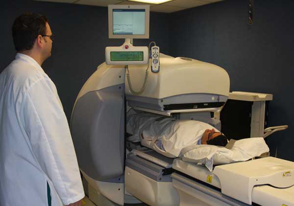
After injection, the radiotracer travels throughout the body to the kidneys, where it gives off energy in the form of gamma rays. This energy is detected by a device called a gamma camera. The camera works with a computer to produce special pictures offering details on both the structure and function of organs and tissues.
What are some common uses of the procedure?
Four types of renal imaging help determine whether the kidneys are working normally or abnormally.
- Renal cortical scintigraphy detects the amount of functioning renal cortical tissue through images taken with a gamma camera approximately two hours after radiopharmaceutical injection.
- Renal perfusion and functional imaging examines blood flow to the kidneys and identifies potential narrowing of the renal arteries. It can also examine whether a renal mass is a focus of benign tissue or a space occupying lesion, such as a cyst or neoplasm. Through a series of images taken over 20 to 30 minutes immediately after radiopharmaceutical injection, it also helps determine how well the kidneys are working.
- Diuretic renal scintigraphy detects kidney blockages or obstruction of urine flow through images taken before and after the introduction of a diuretic, which is used to move urine through the kidneys.
- ACE-inhibitor renal scintigraphy helps determine if the cause of a patient's high blood pressure is coming from the kidneys, due to narrowing of the renal artery or arteries, by comparing kidney images before and after taking a blood pressure medication called an "ACE-inhibitor."
These procedures can be valuable for identifying kidney failure and/or transplant-related complications, as well as discovering kidney-related injuries.
Who interprets the results and how do I get them?
A radiologist or other doctor specially trained in nuclear medicine will interpret the images and send a report to your referring physician.
What are the benefits vs. risks?
Benefits
- The information provided by nuclear renal imaging is unique and often unattainable using other imaging procedures.
- Renal imaging yields useful information needed to make a diagnosis or to determine appropriate treatment, if any.
Risks
- Because nuclear medicine exams use only a small dose of radiotracer, they have a relatively low radiation exposure. This is acceptable for diagnostic exams. Thus, the potential benefits of an exam outweigh the very low radiation risk.
- Doctors have been using nuclear medicine diagnostic procedures for more than six decades. There are no known long-term adverse effects from such low-dose exposure.
- Your doctor always weighs the benefits of nuclear medicine treatment against any risks. Your doctor will discuss the significant risks prior to treatment and give you an opportunity to ask questions.
- Allergic reactions to radiotracers are extremely rare and usually mild. Always tell the nuclear medicine personnel about any allergies you may have. Describe any problems you may have had during previous nuclear medicine exams.
- The radiotracer injection may cause slight pain and redness. This should rapidly resolve.
What are the limitations of renal imaging?
Nuclear renal images cannot reliably differentiate between cysts and tumors.
Nuclear medicine procedures can be time-consuming. You will be informed as to how often and when you will need to return to the nuclear medicine department for further procedures.
The resolution of structures of the body with nuclear medicine may not be as clear as with other imaging techniques, such as CT or MRI. However, nuclear medicine scans are more sensitive than other techniques for a variety of indications, and the functional information gained from nuclear medicine exams is often unobtainable by any other imaging technique.