Last reviewed on September 23, 2024
Abdominal Contrast Enhanced Ultrasound (CEUS)
Abdominal contrast-enhanced ultrasound (CEUS) uses gas-filled microbubbles to better visualize organs and blood vessels within the abdomen and pelvis. This exam may evaluate the liver, spleen, kidneys, pancreas, bowel, and/or bladder.
This procedure requires little to no special preparation. Your doctor will tell you how to prepare, including whether you should not eat or drink beforehand. Wear loose, comfortable clothing. You may need to wear a gown.
What is Abdominal Contrast Enhanced Ultrasound (CEUS)?
CEUS combines ultrasound of the abdomen with a special type of intravenous contrast agent to improve the visualization of blood vessels and organs. CEUS helps the sonographer and the radiologist see abnormalities in the organs or blood vessels. Doctors use this same contrast agent to better assess the heart.
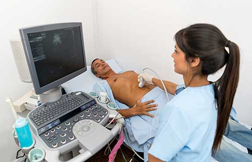
Ultrasound imaging is a noninvasive medical test that helps physicians diagnose and treat medical conditions. It is safe and painless. It produces pictures of the inside of the body using sound waves. Ultrasound imaging is also called sonography. It uses a small probe called a transducer and gel placed directly on the skin. High-frequency sound waves travel from the probe through the gel into the body. The probe collects the sounds that bounce back. A computer uses those sound waves to create an image. Ultrasound exams do not use radiation (x-rays). Because ultrasound captures images in real-time, it can show the structure and movement of the body's internal organs. The images can also show blood flowing through blood vessels.
Abdominal CEUS uses a small amount of contrast material made up of gas-filled microbubbles. The doctor injects this material into the bloodstream through an intravenous (IV) line. Since these tiny microbubbles are similar in size to red blood cells, they pass through the heart and lungs and into arterial circulation. The microbubbles travel via the blood to the organs. Contrast material improves the visualization of organs (and any masses or tumors they may contain) by highlighting blood vessels. This is similar but slightly different than other contrast materials used in CT scans and MRI. Abdominal CEUS is particularly useful for patients who may have kidney failure. This is because the microbubbles can be safely given in all patients, including those with severe kidney disease or those patients on dialysis. Microbubble contrast agents for CEUS are also safe to use in patients who are allergic to the contrast materials used in CT and MRI. CEUS allows you to safely receive more than one contrast material injection during the same exam. This lets your sonographer and radiologist examine multiple lesions or multiple organs during the same session if necessary.
What are some common uses of the procedure?
Doctors use abdominal CEUS to evaluate the:
Doctors also use abdominal CEUS to help diagnose a wide variety of conditions, such as:
- liver lesions discovered during routine ultrasound
- liver lesions that are not well seen on CT or MRI scans
- blood flow abnormalities in the liver
- cirrhosis of the liver
- kidney lesions
- traumatic injuries to the abdomen
- lesions of the spleen
Who interprets the results and how do I get them?
A radiologist, a physician specifically trained to supervise and interpret medical imaging exams, will analyze the images. They will also send a signed report to the doctor who requested the exam. Sometimes, the radiologist will discuss the results with you after the exam. CEUS frequently requires that the radiologist to review the images again while writing their report. This helps them arrive at a definitive diagnosis. In this case, you can get the results from the doctor who ordered the exam once they receive the signed report.
Follow-up exams may be necessary. Your doctor will explain the exact reason why another exam is necessary. Sometimes a follow-up exam is done because a potential abnormality needs further evaluation with additional views or another radiology test. A follow-up exam may also be necessary to monitor a known abnormality over time for any change. Follow-up exams are sometimes the best way to see if a treatment is working or if an abnormality is changing over time.
What are the benefits vs. risks?
Benefits
- An ultrasound exam may occasionally be uncomfortable, but it is rarely painful.
- Ultrasound is widely available, easy-to-use, and less expensive than other imaging methods.
- Ultrasound imaging is extremely safe and does not use any radiation.
- Ultrasound scanning gives a clear picture of many organs that do not show up well on a regular x-ray.
- Abdominal CEUS may eliminate the need for other tests such as CT and/or MRI. This can be especially helpful for patients who have claustrophobia or a hard time laying still.
Risks
- The contrast agents used in abdominal CEUS carry a very small risk of an allergic reaction. This risk is about the same as the risk of allergic reaction to many antibiotics. In fact, it is lower than the risk of allergic reaction to CT and MRI contrast materials.
What are the limitations of Abdominal Contrast Enhanced Ultrasound?
Air can disrupt ultrasound waves. Therefore, a bowel full of air may limit the effectiveness of an ultrasound scan. Ultrasound does not image inside our lungs well for the same reason.
Large or obese patients are more difficult to image by ultrasound. This is because greater amounts of tissue weaken the sound waves as they pass into the body.
Usually only one region of the body can be examined per injection, and only a few areas can be examined in one exam. Because of this, CEUS is not always suitable for looking at multiple organs at once.
Certain lesions and areas of interest may be difficult to image, including lesions less than 1 centimeter, and lesions that are close to the lung or deep within the liver.