Last reviewed on September 09, 2026
How to Prepare for Your X-ray Exam
This is a general description of how to prepare for most X-ray exams. For information about a specific X-ray exam, see the X-ray – Tests and Treatments page.
How to Prepare for Your X-ray Exam
Most x-ray exams require no special preparation.
You may need to remove some clothing and/or change into a gown for the exam. Remove jewelry, removable dental appliances, eyeglasses, and any metal objects or clothing that might interfere with the x-ray images.
Women should always tell their doctor and technologist if they are pregnant. Doctors will not perform many tests during pregnancy to avoid exposing the fetus to radiation. If an x-ray is necessary, the doctor will take precautions to minimize radiation exposure to the baby. See the Radiation Safety page for more information about pregnancy and x-rays.
What does the x-ray equipment look like?
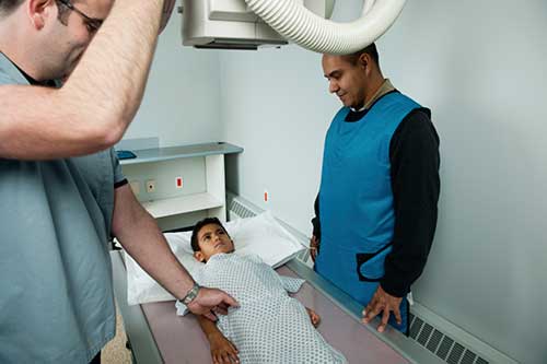
The equipment typically used for an x-ray exam consists of an x-ray tube and a special plate that records the image digitally. The equipment may be arranged with the x-ray tube suspended over a table on which the patient lies. A drawer under the table holds the x-ray film or digital recording plate.
Compact, portable x-ray machines can be taken to the patient in a hospital bed or the emergency room. The x-ray tube is connected to a flexible arm. The technologist extends the arm over the patient and places an x-ray film holder or image recording plate under the patient.
How does x-ray imaging work?
X-rays are a form of radiation like light or radio waves. X-rays pass through most objects, including the body. The technologist carefully aims the x-ray beam at the area of interest. The machine produces a small burst of radiation that passes through your body. The radiation records an image on photographic film or a special detector. Most x-ray exams today are digital, and the images are stored electronically.
Different parts of the body absorb the x-rays in varying degrees. Dense bone absorbs much of the radiation while soft tissue (muscle, fat, and organs) allow more of the x-rays to pass through them. As a result, bones appear white on the x-ray, soft tissue shows up in shades of gray, and air appears black.
On an x-ray, bones absorb much of the radiation and appear white or light gray on the image. Body tissue absorbs little radiation and will appear dark on the image.
Most x-ray images are electronically stored digital files. Your doctor can easily access these stored images to diagnose and manage your condition.
How is the procedure performed?
The radiologic technologist, an individual specially trained to perform radiology exams, will position the patient between the x-ray tube and the image recording plate.
You must hold very still and may need to hold your breath for a few seconds while the radiologic technologist takes the x-ray. This helps reduce the possibility of a blurred image. The technologist will walk behind a wall or into the next room to activate the x-ray machine.
When the exam is complete, the radiologic technologist may ask you to wait until the radiologist confirms they have all the necessary images.
The entire x-ray exam, from positioning to obtaining and verifying the images, is usually completed within 15-30 minutes.
What will I experience during and after the procedure?
An x-ray exam is painless.
You may experience discomfort from the cool temperature in the examination room and the coldness of the recording plate. Individuals with arthritis or injuries to the chest wall, shoulders or arms may have discomfort trying to stay still during the examination. The radiologic technologist will assist you in finding the most comfortable position possible that still ensures diagnostic image quality.