Last reviewed on June 15, 2026
Knee MRI
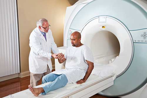
Magnetic resonance imaging (MRI) of the knee uses a powerful magnetic field, radio waves and a computer to produce detailed pictures of the structures within the knee joint. It is typically used to help diagnose or evaluate pain, weakness, swelling or bleeding in and around the joint. Knee MRI does not use ionizing radiation, and it can help determine whether you require surgery.
Tell your doctor about any health problems, recent surgeries or allergies and whether there's a possibility you are pregnant. The magnetic field is not harmful, but it may cause some medical devices to malfunction. Most orthopedic implants pose no risk, but you should always tell the technologist if you have any devices or metal such as shrapnel in your body, especially in or near your brain, spinal cord, heart or eyes. Guidelines about eating and drinking before your exam vary between facilities. Unless you are told otherwise, take your regular medications as usual. Leave jewelry at home and remove any metal piercings. Wear loose, comfortable clothing. You may be asked to wear a gown. If you have claustrophobia or anxiety, you may want to ask your doctor for a mild sedative prior to the exam.
What is a Knee MRI?
MRI of the knee provides detailed images of structures within the knee joint, including bones, cartilage, tendons, ligaments, muscles and blood vessels, from many angles.
Magnetic resonance imaging (MRI) is a noninvasive test doctors use to diagnose medical conditions.
MRI uses a powerful magnetic field, radiofrequency pulses, and a computer to produce detailed pictures of internal body structures. MRI does not use radiation (x-rays).
Detailed MR images allow doctors to examine the body and detect disease.
What are some common uses of the procedure?
In combination with conventional x-rays, MRI is usually the best choice for examining the body's major joints like the knee.
The examination is typically performed to diagnose or evaluate:
- knee pain, weakness, swelling or bleeding in the tissues in and around the joint
- damaged cartilage, meniscus, ligaments or tendons
- sports-related knee injuries, such as sprains and torn ligaments, cartilage, or tendons
- bone fractures that may not be visible on x-rays and other imaging tests
- damage from arthritis
- build-up of fluid in the knee joint
- infections (such as osteomyelitis)
- tumors (primary tumors and metastases) involving bones and joints
- dead bone
- a feeling that your knee is giving away at the joint
- decreased motion of the knee joint
- knee cap injury or pain
- complications related to implanted surgical devices
- pain or trauma following knee surgery
Your doctor may also order an MRI to determine if knee arthroscopy or another surgical procedure is needed, and to monitor your progress after knee surgery.
A special form of MRI called an MR arthrogram injects contrast material into the joint so that the radiologist can get a better look at the knee's structures.
Who interprets the results and how do I get them?
A radiologist, a doctor trained to supervise and interpret radiology exams, will analyze the images. The radiologist will send a signed report to your primary care or referring physician, who will share the results with you.
What are the benefits vs. risks?
Benefits
- MRI is a noninvasive imaging technique that does not involve exposure to radiation.
- MRI has proven valuable in diagnosing a broad range of conditions, including tendon, ligament, muscle, cartilage and bone abnormalities that are not as visible on x-rays or CT scans.
- MRI can help determine which patients with knee injuries require surgery.
- MRI may help diagnose a bone fracture when x-rays and other tests are inconclusive.
- MRI can detect abnormalities that might be obscured by bone with other imaging methods.
- MRI provides a noninvasive alternative to x-ray, angiography and CT for diagnosing problems of the blood vessels.
Risks
- The MRI exam poses almost no risk to the average patient when appropriate safety guidelines are followed.
- If sedation is used, there is a risk of using too much. However, your vital signs will be monitored to minimize this risk.
- The strong magnetic field is not harmful to you. However, it may cause implanted medical devices to malfunction or distort the images.
- Nephrogenic systemic fibrosis is a recognized complication related to injection of gadolinium contrast. It is exceptionally rare with the use of newer gadolinium contrast agents. It usually occurs in patients with serious kidney disease. Your doctor will carefully assess your kidney function before considering a contrast injection.
- There is a very slight risk of an allergic reaction if your exam uses contrast material. Such reactions are usually mild and controlled by medication. If you have an allergic reaction, a doctor will be available for immediate assistance.
- Although there are no known health effects, evidence has shown that very small amounts of gadolinium can remain in the body, particularly the brain, after multiple MRI exams. This is most likely to occur in patients receiving multiple MRI exams over their lifetime for monitoring chronic or high-risk health conditions. The contrast agent is mostly eliminated from the body through the kidneys. If you are a patient in this category, consult with your doctor about the possibility of gadolinium retention, as this effect varies from patient to patient.
- IV contrast manufacturers indicate mothers should not breastfeed their babies for 24-48 hours after contrast material is given. However, the most recent American College of Radiology (ACR) Manual on Contrast Media reports that studies show the amount of contrast absorbed by the infant during breastfeeding is extremely low. For further information please consult the ACR Manual on Contrast Media and its references.
What are the limitations of a knee MRI?
High-quality images are assured only if you are able to remain perfectly still while the images are being recorded. If you are anxious, confused or in severe pain, you may find it difficult to lie still during imaging. Constant coughing and shaking might also interfere with the scan. A bent knee that cannot be extended is also difficult to image.
A person who is very large may not fit into the opening of a conventional MRI machine.
The presence of an implant or other metallic object sometimes makes it difficult to obtain clear images and patient movement can have the same effect. In some cases, metal artifact reduction imaging is performed in patients who have metallic surgical implants at the knee and require MR imaging.
Present data show no convincing evidence that non contrast MRI harms the fetus of a pregnant woman. However, if the need for the exam is not time sensitive your doctor may delay the exam until after delivery. MRI gadolinium contrast agents are generally avoided during pregnancy except in very specific circumstances. Your doctor will discuss the benefits and risks of any MRI procedure with you. Doctors may perform MRI after the first trimester to assess the fetus for findings that are not fully evaluated by ultrasound.
An MRI exam typically costs more and may take more time than other imaging exams. Talk to your insurance provider if you have concerns about the cost of MRI.