Last reviewed on June 15, 2026
CT Angiography (CTA)
Computed tomography angiography (CTA) uses an injection of contrast material into your blood vessels and CT scanning to help diagnose and evaluate blood vessel disease or related conditions, such as aneurysms or blockages. CTA is typically performed in a radiology department or an outpatient imaging center.
Tell your doctor if there is a possibility you are pregnant and discuss any recent illnesses, medical conditions, medications you are taking, and allergies. You will be instructed not to eat or drink anything several hours beforehand. If you have a known allergy to contrast material, your doctor may prescribe medications to take before the CTA exam to reduce the risk of an allergic reaction. Leave jewelry at home and wear loose, comfortable clothing. You may be asked to wear a gown. If you are breastfeeding, talk to your doctor about how to proceed.
What is CT Angiography?
Doctors use angiography to diagnose and treat blood vessel diseases and conditions. Angiography exams produce pictures of major blood vessels throughout the body. Most exams use contrast material.
Doctors perform angiography using:
- x-rays with catheters
- computed tomography (CT)
- magnetic resonance imaging (MRI)
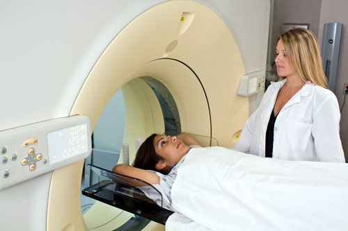
CT angiography uses a CT scanner to produce detailed images of both blood vessels and tissues in various parts of the body. During the exam, contrast material is injected through a small catheter placed in a vein of the arm. A radiologic technologist will capture high-resolution CT images while the contrast material flows through the blood vessels.
What are some common uses of the procedure?
CT angiography is helpful in examining blood vessels and the organs supplied by them in various body parts, including:
- brain
- neck
- heart
- chest
- abdomen (such as the kidneys and liver)
- pelvis
- legs and feet
- arms and hands
Physicians use CT angiography to diagnose and evaluate many diseases of blood vessels and related conditions such as:
- aneurysms
- blockages
- blood clots
- congenital (birth-related) abnormalities of the cardiovascular system, including the heart
- disorganized blood vessels, such as vascular malformations
- injury
- tumors
- vessel rupture or tears
Also, physicians use CT angiography to check blood vessels before or following surgery to:
- identify abnormalities, such as aneurysms, in the aorta, both in the chest and abdomen, or in other arteries.
- detect atherosclerotic (plaque) disease in the carotid artery of the neck, which may limit blood flow to the brain and cause a stroke.
- identify a arteriovenous malformation inside the brain or elsewhere.
- detect plaque disease that has narrowed the arteries to the legs and help prepare for angioplasty/stent placement or surgery.
- detect disease in the arteries to the kidneys or visualize blood flow to help prepare for a kidney transplant or stent placement.
- guide interventional radiologists and surgeons making repairs to diseased blood vessels, such as implanting stents or evaluating a stent after implantation.
- detect injury to one or more arteries in the neck, chest, abdomen, pelvis, or limbs following trauma.
- evaluate arteries feeding a tumor prior to surgery or other procedures such as chemoembolization or selective internal radiation therapy.
- identify dissection or splitting in the aorta in the chest or abdomen or its major branches.
- show the extent and severity of coronary artery disease and its effects and plan for an intervention, such as a coronary bypass and stenting.
- examine pulmonary arteries in the lungs to detect pulmonary embolism (blood clots, such as those traveling from leg veins) or pulmonary arteriovenous malformations.
- look at congenital abnormalities in blood vessels, especially arteries in children (e.g., malformations in the heart or other blood vessels due to congenital heart disease).
- evaluate stenosis and obstructions of vessels.
Who interprets the results and how do I get them?
A radiologist, a doctor specially trained to supervise and interpret radiology exams, will analyze the images. The radiologist will send an official report to the doctor who ordered the exam.
You may need a follow-up exam. If so, your doctor will explain why. Sometimes a follow-up exam further evaluates a potential issue with more views or a special imaging technique. It may also see if there has been any change in an issue over time. Follow-up exams are often the best way to see if treatment is working or if a problem needs attention.
What are the benefits vs. risks?
Benefits
- Angiography may eliminate the need for surgery. If surgery remains necessary, it can be performed more accurately.
- CT angiography is fast, non-invasive and may have fewer complications compared to conventional angiography.
- CT angiography may provide more precise anatomical details than other angiography exams such as conventional catheter angiography and magnetic resonance imaging (MRI).
- For CT Angiography, there is no need for sedation or general anesthesia.
- CT angiography of the heart helps detect blocked coronary arteries.
- CT angiography may also cost less than catheter angiography.
- No radiation remains in a patient's body after a CT exam.
- The x-rays used for CT scanning should have no immediate side effects.
Risks
Most patients complete CT angiography with no adverse events.
- There is always a slight chance of cancer from excessive exposure to radiation. However, the benefit of an accurate diagnosis far outweighs the risk involved with CT scanning.
- If you have a history of allergy to x-ray contrast material, your doctor may advise you to take special precautionary medication, such as a steroid, for a few hours or the day before CT angiography to lessen the chances of an allergic reaction. Another option is to undergo a different exam that does not require iodinated contrast material.
- In patients who are at risk for kidney failure and who already have borderline kidney function, administering iodinated contrast material could potentially further damage kidney function. Check with your referring doctor and radiologist to obtain more information regarding this risk.
- If a large amount of x-ray contrast material leaks out from the vein being injected and spreads under the skin where the IV is placed, it may damage the skin, blood vessels and nerves. If you feel any pain or tingling sensation in this area during or immediately after the contrast material injection, you should immediately inform the nurse/technologist.
- Women should always tell their doctor and x-ray or CT technologist if there is any chance they are pregnant. See the Radiation Safety page for more information about pregnancy and x-rays.
- IV contrast manufacturers indicate mothers should not breastfeed their babies for 24-48 hours after contrast material is given. However, the most recent American College of Radiology (ACR) Manual on Contrast Media reports that studies show the amount of contrast absorbed by the infant during breastfeeding is extremely low. For further information please consult the ACR Manual on Contrast Media and its references.
- The risk of severe allergic reaction to contrast materials that contain iodine is rare, and hospitals are well-equipped to deal with these reactions.
A Word About Minimizing Radiation Exposure
Doctors take special care during x-ray exams to use the lowest radiation dose possible while producing the best images for evaluation. National and international radiation protection organizations continually review and update the technique standards radiology professionals use.
Modern x-ray systems minimize stray (scatter) radiation by using controlled x-ray beams and dose control methods. This ensures that the areas of your body not being imaged receive minimal radiation exposure.
Every effort will be made to reduce radiation while performing CT angiography, including tailoring the scan parameters specific to your body type. The scanning area will also be limited to the organ of interest to avoid unnecessary radiation to other body parts.
What are the limitations of CT Angiography?
A person who is very large may not fit into the opening of a conventional CT scanner. Or, they may be over the weight limit—usually 450 pounds—for the moving table.
Some patients with impaired renal function or who previously exhibited a severe allergic reaction to iodinated contrast material may not be candidates for CT angiography.