Last reviewed on May 01, 2023
Ultrasound - Thyroid
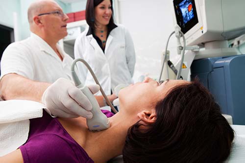
Thyroid ultrasound uses sound waves to produce pictures of the thyroid gland within the neck. It does not use ionizing radiation and is commonly used to evaluate lumps or nodules found during a routine physical or other imaging exam.
This procedure requires little to no special preparation. Leave jewelry at home and wear loose, comfortable clothing. You may be asked to wear a gown.
What is an Ultrasound of the Thyroid?
Ultrasound imaging is a noninvasive medical test that helps physicians diagnose and treat medical conditions. It is safe and painless. It produces pictures of the inside of the body using sound waves. Ultrasound imaging is also called sonography. It uses a small probe called a transducer and gel placed directly on the skin. High-frequency sound waves travel from the probe through the gel into the body. The probe collects the sounds that bounce back. A computer uses those sound waves to create an image. Ultrasound exams do not use radiation (x-rays). Because ultrasound captures images in real-time, it can show the structure and movement of the body's internal organs. The images can also show blood flowing through blood vessels.
An ultrasound of the thyroid produces pictures of the thyroid gland and the adjacent structures in the neck. The thyroid gland is located in front of the neck just above the collar bones and is shaped like a butterfly, with one lobe on either side of the neck connected by a narrow band of tissue called the thyroid isthmus. It is one of nine endocrine glands located throughout the body that make and send hormones into the bloodstream.
The thyroid gland makes the thyroid hormone, which helps to regulate a variety of body functions including how fast the heart beats. It is very common for patchy areas or nodules to develop in the thyroid that may or may not be felt on the skin surface. About five to 10 percent of adults will have lumps in their thyroid that a doctor can identify on an exam. These are called palpable nodules. Ultrasound is very sensitive and shows many nodules that cannot be felt. In some age groups, nodules are seen on ultrasound in as many as 70 percent of adults. The vast majority of these are benign regions of thyroid tissue that pose no health risk. The minority of these are true tumors of the thyroid and may require further diagnosis or treatment.
What are some common uses of the procedure?
An ultrasound of the thyroid is typically used:
- to determine if a lump in the neck is arising from the thyroid or an adjacent structure
- to analyze the appearance of thyroid nodules and determine if they are the more common benign nodule or if the nodule has features that require a biopsy. If biopsy is required, ultrasound-guided fine needle aspiration can help improve accuracy of the biopsy.
- to look for additional nodules in patients with one or more nodules felt on physical exam
- to see if a thyroid nodule has substantially grown over time
Because ultrasound provides real-time images, doctors may use it to guide procedures, including needle biopsies. Biopsies use needles to extract tissue samples for lab testing. Doctors also use ultrasound to guide insertion of a catheter or other drainage device. This helps assure safe and accurate placement.
Who interprets the results and how do I get them?
A radiologist, a doctor trained to supervise and interpret radiology exams, will analyze the images. The radiologist will send a signed report to the doctor who requested the exam. Your doctor will then share the results with you. In some cases, the radiologist may discuss results with you after the exam.
You may need a follow-up exam. If so, your doctor will explain why. Sometimes a follow-up exam further evaluates a potential issue with more views or a special imaging technique. It may also see if there has been any change in an issue over time. Follow-up exams are often the best way to see if treatment is working or if a problem needs attention.
What are the benefits vs. risks?
Benefits
- Most ultrasound scanning is noninvasive (no needles or injections).
- Occasionally, an ultrasound exam may be temporarily uncomfortable, but it should not be painful.
- Ultrasound is widely available, easy to use, and less expensive than most other imaging methods.
- Ultrasound imaging is extremely safe and does not use radiation.
- Ultrasound scanning gives a clear picture of soft tissues that do not show up well on x-ray images.
- Ultrasound provides real-time imaging. This makes it a good tool for guiding minimally invasive procedures such as needle biopsies and fluid aspiration.
Risks
- Standard diagnostic ultrasound has no known harmful effects on humans.
What are the limitations of an Ultrasound of the Thyroid?
If one or more nodules are detected within the thyroid gland, the radiologist will examine the features of the nodules. Some features are strongly suggestive that a nodule is benign in nature, and some raise concern that the nodule may be a true tumor. In other cases, the radiologist cannot distinguish between benign and malignant lumps with complete certainty. A fine needle biopsy and review of tissue under a microscope may be recommended for further evaluation, but in some cases surveillance and a repeat sonogram after a few months looking for stability may suffice.
It is not possible to determine thyroid function—that is, whether the thyroid gland is underactive, overactive, or normal—with ultrasound. For that determination, your doctor may order a blood test or a radioactive iodine uptake test.
Additional Information and Resources
RTAnswers.org: Radiation Therapy for Head and Neck Cancer