Last reviewed on June 15, 2026
Chest MRI
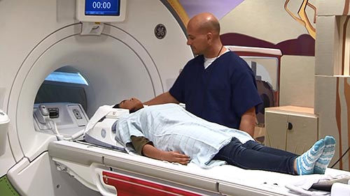
Magnetic resonance imaging (MRI) of the chest uses a powerful magnetic field, radio waves and a computer to produce detailed pictures of the structures within the chest. It is primarily used to characterize abnormalities found on other imaging tests such as Chest CT. It is also used to assess the anatomy and function of the heart and its blood flow.
Tell your doctor about any health problems, recent surgeries, devices or metal in your body, allergies and whether there is a possibility you are pregnant. The magnetic field is not harmful to you, but it may cause some medical devices to malfunction. Most orthopedic implants pose no risk. If you have manufacturer information about your implanted device, please bring it with you for your MRI appointment. Guidelines about eating and drinking before your exam vary between facilities. Unless you are told otherwise, take your regular medications as usual. Leave jewelry at home. You will need to change into a gown for the exam. If you have claustrophobia or anxiety, you may want to ask your doctor for a mild sedative prior to the exam.
What is MRI of the Chest?
Magnetic resonance imaging (MRI) is a noninvasive test doctors use to diagnose medical conditions.
MRI uses a powerful magnetic field, radiofrequency pulses, and a computer to produce detailed pictures of internal body structures. MRI does not use radiation (x-rays).
Detailed MR images allow doctors to examine the body and detect disease.
MRI of the chest gives detailed pictures of structures within the chest cavity, including the mediastinum, chest wall, pleura, heart and vessels, from almost any angle. MRI also provides movie-like sequential imaging of the cardiovascular system that is important to assess the health and function of these structures (heart, valves, great vessels, etc.).
What are some common uses of the procedure?
MR imaging of the chest is performed to:
- characterize abnormal masses, which either cannot be assessed adequately with other imaging modalities (typically CT)
- determine tumor size, extent, and the degree of spread to adjacent structures.
- assess the anatomy and function of the heart and its component structures (valves, etc.).
- assess myocardial perfusion (blood flow to the heart) and myocardial infarct (scar in the heart muscle due to prior obstruction of blood flow).
- determine blood flow dynamics in the vessels and heart chambers.
- display lymph nodes and blood vessels, including vascular and lymphatic malformations of the chest.
- assess disorders of the chest bones (vertebrae, ribs, and sternum) and chest wall soft tissue (muscles and fat).
- assess for pericardial (thin sac around the heart) disease.
- characterize mediastinal or pleural lesions seen by other imaging modalities, such as chest x-ray or CT.
A special form of MRI called magnetic resonance angiography (MRA) is helpful to assess the vessels of the chest cavity (arteries and veins). MRA can also demonstrate an abnormal ballooning out of the wall of an artery (aneurysm), blockage of a vessel (thrombosis), or a torn inner lining of an artery (dissection). See the MRA page for more information.
Who interprets the results and how do I get them?
A radiologist, a doctor trained to supervise and interpret radiology exams, will analyze the images. The radiologist will send a signed report to your primary care or referring physician, who will share the results with you.
You may need a follow-up exam. If so, your doctor will explain why. Sometimes a follow-up exam further evaluates a potential issue with more views or a special imaging technique. It may also see if there has been any change in an issue over time. Follow-up exams are often the best way to see if treatment is working or if a problem needs attention.
What are the benefits vs. risks?
Benefits
- MRI is a noninvasive imaging technique that does not involve exposure to radiation.
- MR images of the heart and vascular structures are often clearer and more detailed than with other imaging methods. This detail makes MRI an invaluable tool in early diagnosis and evaluation of cardiovascular conditions.
- MRI has proven valuable in diagnosing a broad range of conditions, including heart and vascular disease, heart valve abnormalities, bone, and other soft tissue abnormalities of the chest. MRI is also useful for staging tumors.
- MRI can help physicians evaluate both the structure of an organ and how it is working.
- MRI can detect abnormalities that might be obscured by bone with other imaging methods.
- The MRI gadolinium contrast material is less likely to cause an allergic reaction than the iodine-based contrast materials used for x-rays and CT scanning.
- MRI of the chest is often more informative than other imaging procedures for differentiating and characterizing soft tissues, except for lung abnormalities where Chest CT is a preferred imaging test.
- MR imaging can assess blood flow without risking the side effects of conventional (catheter) angiography.
Risks
- The MRI exam poses almost no risk to the average patient when appropriate safety guidelines are followed.
- If sedation is used, there is a risk of using too much. However, your vital signs will be monitored to minimize this risk.
- The strong magnetic field is not harmful to you. However, it may cause implanted medical devices to malfunction or distort the images.
- Nephrogenic systemic fibrosis is a recognized complication related to injection of gadolinium contrast. It is exceptionally rare with the use of newer gadolinium contrast agents. It usually occurs in patients with serious kidney disease. Your doctor will carefully assess your kidney function before considering a contrast injection.
- There is a very slight risk of an allergic reaction if your exam uses contrast material. Such reactions are usually mild and controlled by medication. If you have an allergic reaction, a doctor will be available for immediate assistance.
- Although there are no known health effects, evidence has shown that very small amounts of gadolinium can remain in the body, particularly the brain, after multiple MRI exams. This is most likely to occur in patients receiving multiple MRI exams over their lifetime for monitoring chronic or high-risk health conditions. The contrast agent is mostly eliminated from the body through the kidneys. If you are a patient in this category, consult with your doctor about the possibility of gadolinium retention, as this effect varies from patient to patient.
- IV contrast manufacturers indicate mothers should not breastfeed their babies for 24-48 hours after contrast material is given. However, the most recent American College of Radiology (ACR) Manual on Contrast Media reports that studies show the amount of contrast absorbed by the infant during breastfeeding is extremely low. For further information please consult the ACR Manual on Contrast Media and its references.
What are the limitations of MRI of the Chest?
High-quality images depend on your ability to remain perfectly still and follow breath-holding instructions while the images are being recorded. If you are anxious, confused or in severe pain, you may find it difficult to lie still during imaging.
A person who is very large may not fit into certain types of MRI machines. There are weight limits on the scanners.
Implants and other metallic objects can make it difficult to obtain clear images. Patient movement can have the same effect.
A very irregular heartbeat may affect the quality of images. This is because some techniques time the imaging based on the electrical activity of the heart.
MRI is generally not recommended for seriously injured patients. However, this decision is based on clinical judgment. This is because traction devices and life support equipment may distort the MR images. As a result, they must be kept away from the area to be imaged. Some trauma patients, however, may need MRI.
Present data show no convincing evidence that non contrast MRI harms the fetus of a pregnant woman. However, if the need for the exam is not time sensitive your doctor may delay the exam until after delivery. MRI gadolinium contrast agents are generally avoided during pregnancy except in very specific circumstances. Your doctor will discuss the benefits and risks of any MRI procedure with you. Doctors may perform MRI after the first trimester to assess the fetus for findings that are not fully evaluated by ultrasound.
An MRI exam typically costs more and may take more time than other imaging exams. Talk to your insurance provider if you have concerns about the cost of MRI.
MRI of the chest takes more time than an x-ray or CT exam. Because of the length of time an MRI takes to complete, many young children and infants require sedation to hold still for the exam.