skip to main content
RadInfo Logo Home

MR Enterography

Magnetic resonance (MR) enterography is an imaging test which produces detailed pictures of your small intestine. It may help your doctor diagnose inflammation, bleeding, obstructions and other problems. It is noninvasive and does not use ionizing radiation.

The exam uses a magnetic field to create detailed images of your organs. A computer analyzes the images. Before the exam, oral and intravenous contrast material are administered to highlight the small intestine. A drug may also be administered to decrease movement of the bowel which can interfere with the images. Tell your doctor about any health problems, recent surgeries or allergies and whether there's a possibility you are pregnant. The magnetic field is not harmful, but it may cause some medical devices to malfunction. Most orthopedic implants pose no risk, but you should always tell the technologist if you have any devices or metal in your body.

Guidelines about eating and drinking before your exam vary between facilities. Unless you are told otherwise, take your regular medications as usual. Leave jewelry at home and wear loose, comfortable clothing. You may be asked to wear a gown. If you have claustrophobia or anxiety, you may want to ask your doctor for a mild sedative prior to the exam.

What is MR Enterography?

MR enterography is a special type of magnetic resonance imaging (MRI) performed with a contrast material to produce detailed images of the small intestine.

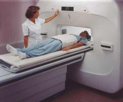
Magnetic resonance imaging (MRI) is a noninvasive test doctors use to diagnose medical conditions.

MRI uses a powerful magnetic field, radiofrequency pulses, and a computer to produce detailed pictures of internal body structures. MRI does not use radiation (x-rays).

Detailed MR images allow doctors to examine the body and detect disease.

What are some common uses of the procedure?

Physicians use MR enterography to identify and locate:

How should I prepare?

You will need to change into a hospital gown. This is to prevent artifacts appearing on the final images and to comply with safety regulations related to the strong magnetic field.

Guidelines about eating and drinking before an MRI vary between specific exams and facilities. Take food and medications as usual unless your doctor tells you otherwise.

Some MRI exams use an injection of contrast material. The doctor may ask if you have asthma or allergies to contrast material, drugs, food, or the environment. MRI exams commonly use a contrast material called gadolinium. Doctors can use gadolinium in patients who are allergic to iodine contrast. A patient is much less likely to be allergic to gadolinium than to iodine contrast. However, even if the patient has a known allergy to gadolinium, it may be possible to use it after appropriate pre-medication. For more information on allergic reactions to gadolinium contrast, please consult the ACR Manual on Contrast Media.

Tell the technologist or radiologist if you have any serious health problems or recent surgeries. Some conditions, such as severe kidney disease, may mean that you cannot safely receive gadolinium. You may need a blood test to confirm your kidneys are functioning normally.

Women should always tell their doctor and technologist if they are pregnant. MRI has been used since the 1980s with no reports of any ill effects on pregnant women or their unborn babies. However, the baby will be in a strong magnetic field. Therefore, pregnant women should not have an MRI in the first trimester unless the benefit of the exam clearly outweighs any potential risks. Pregnant women should not receive gadolinium contrast unless absolutely necessary. See the MRI Safety During Pregnancy page for more information about pregnancy and MRI.

If you have claustrophobia (fear of enclosed spaces) or anxiety, ask your doctor to prescribe a mild sedative prior to the date of your exam.

Infants and young children often require sedation or anesthesia to complete an MRI exam without moving. This depends on the child's age, intellectual development, and the type of exam. Sedation can be provided at many facilities. A specialist in pediatric sedation or anesthesia should be available during the exam for your child's safety. You will be told how to prepare your child.

Some facilities may have personnel who work with children to help avoid the need for sedation or anesthesia. They may prepare children by showing them a model MRI scanner and playing the noises they might hear during the exam. They also answer any questions and explain the procedure to relieve anxiety. Some facilities also provide goggles or headsets so the child can watch a movie during the exam. This helps the child stay still and allows for good quality images.

Leave all jewelry and other accessories at home or remove them prior to the MRI scan. Metal and electronic items are not allowed in the exam room. They can interfere with the magnetic field of the MRI unit, cause burns, or become harmful projectiles. These items include:

  • jewelry, watches, credit cards, and hearing aids, all of which can be damaged
  • pins, hairpins, metal zippers, and similar metallic items, which can distort MRI images
  • removable dental work
  • pens, pocketknives, and eyeglasses
  • body piercings
  • mobile phones, electronic watches, and tracking devices.

In most cases, an MRI exam is safe for patients with metal implants, except for a few types. People with the following implants may not be scanned and should not enter the MRI scanning area without first being evaluated for safety:

Tell the technologist if you have medical or electronic devices in your body. These devices may interfere with the exam or pose a risk. Many implanted devices will have a pamphlet explaining the MRI risks for that device. If you have the pamphlet, bring it to the attention of the scheduler before the exam. MRI cannot be performed without confirmation and documentation of the type of implant and MRI compatibility. You should also bring any pamphlet to your exam in case the radiologist or technologist has any questions.

If there is any question, an x-ray can detect and identify any metal objects. Metal objects used in orthopedic surgery generally pose no risk during MRI. However, a recently placed artificial joint may require the use of a different imaging exam.

Tell the technologist or radiologist about any shrapnel, bullets, or other metal that may be in your body. Foreign bodies near and especially lodged in the eyes are very important because they may move or heat up during the scan and cause blindness. Dyes used in tattoos may contain iron and could heat up during an MRI scan. This is rare. The magnetic field will usually not affect tooth fillings, braces, eyeshadows, and other cosmetics. However, these items may distort images of the facial area or brain. Tell the radiologist about them.

Anyone accompanying a patient into the exam room must also undergo screening for metal objects and implanted devices.

What does the equipment look like?

The traditional MRI unit is a large cylinder-shaped tube surrounded by a circular magnet. You will lie on a table that slides into a tunnel towards the center of the magnet.

Some MRI units, called short-bore systems, are designed so that the magnet does not completely surround you. Some newer MRI machines have a larger diameter bore, which can be more comfortable for larger patients or those with claustrophobia. "Open" MRI units are open on the sides. They are especially helpful for examining larger patients or those with claustrophobia. Open MRI units can provide high quality images for many types of exams. Open MRI may not be used for certain exams. For more information, consult your radiologist.

How does the procedure work?

Unlike x-ray and computed tomography (CT) exams, MRI does not use radiation. Instead, radio waves re-align hydrogen atoms that naturally exist within the body. This does not cause any chemical changes in the tissues. As the hydrogen atoms return to their usual alignment, they emit different amounts of energy depending on the type of tissue they are in. The scanner captures this energy and creates a picture using this information.

In most MRI units, the magnetic field is produced by passing an electric current through wire coils. Other coils are inside the machine and, in some cases, are placed around the part of the body being imaged. These coils send and receive radio waves, producing signals that are detected by the machine. The electric current does not come into contact with the patient.

A computer processes the signals and creates a series of images, each of which shows a thin slice of the body. The radiologist can study these images from different angles.

MRI is often able to tell the difference between diseased tissue and normal tissue better than x-ray, CT, and ultrasound.

How is the procedure performed?

MRI exams may be done on an outpatient basis.

Prior to the procedure, you will be asked to drink several glasses of a water solution mixed with a contrast material.

The technologist will position you on the moveable exam table. They may use straps and bolsters to help you stay still and maintain your position.

The technologist may place devices that contain coils capable of sending and receiving radio waves around or next to the area of the body under examination.

MRI exams generally include multiple runs (sequences), some of which may last several minutes. Each run will create a different set of noises.

If your exam uses a contrast material, a doctor, nurse, or technologist will insert an intravenous catheter (IV line) into a vein in your hand or arm. They will use this IV to inject the contrast material.

You will be placed into the magnet of the MRI unit. The technologist will perform the exam while working at a computer outside of the room. You will be able to talk to the technologist via an intercom.

If your exam uses a contrast material, the technologist will inject it into the intravenous line (IV) after an initial series of scans. They will take more images during or following the injection.

When the exam is complete, the technologist may ask you to wait while the radiologist checks the images in case more are needed.

The technologist will remove your IV line after the exam is over and place a small dressing over the insertion site.

What will I experience during and after the procedure?

Most MRI exams are painless. However, some patients find it uncomfortable to remain still. Others may feel closed-in (claustrophobic) while in the MRI scanner. The scanner can be noisy.

It is normal for the area of your body being imaged to feel slightly warm. If it bothers you, tell the radiologist or technologist. It is important that you remain perfectly still while the images are being taken. This is typically only a few seconds to a few minutes at a time. You will know when images are being recorded because you will hear and feel loud tapping or thumping sounds. The coils that generate the radio waves make these sounds when they are activated. You will be provided with earplugs or headphones to reduce the noise made by the scanner. You may be able to relax between imaging sequences. However, you will need to keep the same position as much as possible without moving.

You will usually be alone in the exam room. However, the technologist will be able to see, hear, and speak with you at all times using a two-way intercom. They will give you a “squeeze-ball” that alerts the technologist that you need attention right away. Many facilities allow a friend or parent to stay in the room if they have also been screened for safety.

Children will be given appropriately sized earplugs or headphones during the exam. Music may be played through the headphones to help pass the time. MRI scanners are air-conditioned and well-lit.

In some cases, IV injection of contrast material may be given before the images are obtained. The IV needle may cause you some discomfort and you may experience some bruising. There is also a very small chance of skin irritation at the site of the IV tube insertion. Some patients may have a temporary metallic taste in their mouth after the contrast injection.

If you do not require sedation, no recovery period is necessary. You may resume your usual activities and normal diet immediately after the exam. On very rare occasions, a few patients experience side effects from the contrast material. These may include nausea, headache, and pain at the site of injection. It is very rare that patients experience hives, itchy eyes, or other allergic reactions to the contrast material. If you have allergic symptoms, tell the technologist. A radiologist or other doctor will be available for immediate assistance.

Manufacturers of intravenous contrast indicate mothers should not breastfeed their babies for 24-48 hours after contrast medium is given. However, both the American College of Radiology (ACR) and the European Society of Urogenital Radiology note that the available data suggest that it is safe to continue breastfeeding after receiving intravenous contrast. For further information please consult the ACR Manual on Contrast Media and its references.

You will be asked to drink the oral contrast material both quickly and steadily so the liquid contrast fills your entire small bowel.

The contrast material may cause nausea, diarrhea and abdominal cramps. You should tell your doctor if these mild side effects become severe or do not go away.

See the Contrast Materials page for more information.

Who interprets the results and how do I get them?

A radiologist, a doctor trained to supervise and interpret radiology exams, will analyze the images. The radiologist will send a signed report to your primary care or referring physician, who will share the results with you.

What are the benefits vs. risks?

Benefits

  • MRI is a noninvasive imaging technique that does not involve exposure to radiation.
  • MRI can detect abnormalities that might be obscured by bone with other imaging methods.
  • The MRI gadolinium contrast material is less likely to cause an allergic reaction than the iodine-based contrast materials used for x-rays and CT scanning.
  • MR enterography is a complementary imaging examination that helps identify areas of bowel inflammation due to such diseases as Crohn's.
  • Because MR enterography does not involve ionizing radiation, the procedure may be preferred for the evaluation of young patients with inflammatory bowel disease who may undergo multiple exams throughout their life.
  • MR enterography may eliminate the need for video capsule endoscopy (VCE).

Risks

  • The MRI exam poses almost no risk to the average patient when appropriate safety guidelines are followed.
  • If sedation is used, there is a risk of using too much. However, your vital signs will be monitored to minimize this risk.
  • The strong magnetic field is not harmful to you. However, it may cause implanted medical devices to malfunction or distort the images.
  • Nephrogenic systemic fibrosis is a recognized complication related to injection of gadolinium contrast. It is exceptionally rare with the use of newer gadolinium contrast agents. It usually occurs in patients with serious kidney disease. Your doctor will carefully assess your kidney function before considering a contrast injection.
  • There is a very slight risk of an allergic reaction if your exam uses contrast material. Such reactions are usually mild and controlled by medication. If you have an allergic reaction, a doctor will be available for immediate assistance.
  • Although there are no known health effects, evidence has shown that very small amounts of gadolinium can remain in the body, particularly the brain, after multiple MRI exams. This is most likely to occur in patients receiving multiple MRI exams over their lifetime for monitoring chronic or high-risk health conditions. The contrast agent is mostly eliminated from the body through the kidneys. If you are a patient in this category, consult with your doctor about the possibility of gadolinium retention, as this effect varies from patient to patient.

What are the limitations of MR Enterography?

High-quality images depend on your ability to remain perfectly still and follow breath-holding instructions while the images are being recorded. If you are anxious, confused or in severe pain, you may find it difficult to lie still during imaging.

A person who is very large may not fit into certain types of MRI machines. There are weight limits on the scanners.

Implants and other metallic objects can make it difficult to obtain clear images. Patient movement can have the same effect.

A very irregular heartbeat may affect the quality of images. This is because some techniques time the imaging based on the electrical activity of the heart.

Present data show no convincing evidence that non contrast MRI harms the fetus of a pregnant woman. However, if the need for the exam is not time sensitive your doctor may delay the exam until after delivery. MRI gadolinium contrast agents are generally avoided during pregnancy except in very specific circumstances. Your doctor will discuss the benefits and risks of any MRI procedure with you. Doctors may perform MRI after the first trimester to assess the fetus for findings that are not fully evaluated by ultrasound.

For optimal results the patient should consume the entire dose of oral contrast material, remain still and follow the breathing instructions of an MR technologist. Moreover, results may be compromised if the patient is not able to receive the intravenous contrast material (gadolinium).

MR enterography takes longer to perform (30 to 45 minutes) than CT enterography (two to four minutes).

This page was reviewed on May 01, 2023

Images

Magnetic Resonance Imaging (MRI)equipment. This is an example of an 'open' MRI unit. Open MRI models are designed to alleviate patient claustrophobia. View full size with caption

Pediatric Content

Pediatric
Some imaging tests and treatments have special pediatric considerations. The hand denotes child-specific content.

Sponsored By

Please note

RadiologyInfo.org is not a medical facility. Please contact your physician with specific medical questions or for a referral to a radiologist or other physician. To locate a medical imaging or radiation oncology provider in your community, you can search the ACR-accredited facilities database.

This website does not provide cost information. The costs for specific medical imaging tests, treatments and procedures may vary by geographic region. Discuss the fees associated with your prescribed procedure with your doctor, the medical facility staff and/or your insurance provider to get a better understanding of the possible charges you will incur.

Web page review process: This Web page is reviewed regularly by a physician with expertise in the medical area presented and is further reviewed by committees from the Radiological Society of North America (RSNA) and the American College of Radiology (ACR), comprising physicians with expertise in several radiologic areas.

Outside links: For the convenience of our users, RadiologyInfo.org provides links to relevant websites. RadiologyInfo.org, RSNA and ACR are not responsible for the content contained on the web pages found at these links.