Last reviewed on June 15, 2026
Brain MRI
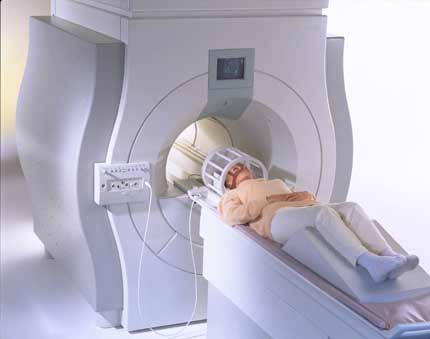
A brain MRI, also called a head MRI, uses a powerful magnetic field, radio waves and a computer to produce pictures of the brain. The pictures produced are clearer and more detailed than other imaging methods. Magnetic resonance imaging (MRI) does not use ionizing radiation and may require an injection of a contrast material called gadolinium.
Tell your doctor about any health problems, recent surgeries or allergies and whether there's a possibility you are pregnant. The magnetic field is not harmful, but it may cause some medical devices to malfunction. Most orthopedic implants pose no risk, but you should always tell the radiologic technologist if you have any devices or metal in your body. Guidelines about eating and drinking before your exam vary between facilities. Unless you are told otherwise, take your regular medications as usual. Leave jewelry at home and wear loose, comfortable clothing. You may be asked to wear a gown. If you have claustrophobia or anxiety, you may want to ask your doctor for a mild sedative prior to the exam.
What is a brain MRI?
Magnetic resonance imaging (MRI) uses a powerful magnetic field, radiofrequency pulses, and a computer to produce detailed pictures of internal body structures. MRI does not use radiation (x-rays).
Detailed MR images allow doctors to examine the body and detect disease. Currently, MRI is the best imaging test for many brain disorders.
What are some common uses of brain MRI?
Doctors use Brain MRI to investigate sudden onset or long-standing symptoms. It can help diagnose conditions such as:
- stroke and stroke damage
- changes associated with aging (brain volume loss or signal changes in brain tissue)
- brain damage or bleeding from traumatic injury
- loss of body movement control (ataxia)
- brain tumors
- developmental anomalies
- hydrocephalus — dilatation of fluid spaces within the brain (ventricles)
- causes of epilepsy (seizure)
- infections
- certain chronic conditions, such as multiple sclerosis
- eye and inner ear disorders
- pituitary gland disorders
Who interprets the results and how do I get them?
A radiologist, a doctor trained to supervise and interpret radiology exams, will analyze the images. The radiologist will send a signed report to your ordering provider, who will share the results with you.
You may need a follow-up exam. If so, your doctor will explain why. Sometimes a follow-up exam further evaluates a potential issue with more views or a special imaging technique. It may also see if there has been any change in an issue over time. Follow-up exams are often the best way to see if treatment is working or if a problem needs attention.
What are the benefits vs. risks?
Benefits
- MRI is a noninvasive imaging technique that does not involve exposure to radiation.
- MRI can help physicians evaluate the structures of the brain and can also provide functional information in selected cases.
- MR images of the brain and other cranial structures are clearer and more detailed than with other imaging methods. This detail makes MRI an invaluable tool in early diagnosis and evaluation of many conditions, including tumors.
- MRI can detect abnormalities that might be obscured by bone with other imaging methods.
- A variant called MR angiography (MRA) provides detailed images of blood vessels in the brain—often without the need for contrast material. See the MRA page for more information.
Risks
- The MRI exam poses almost no risk to the average patient when appropriate safety guidelines are followed.
- The strong magnetic field is not harmful to you. However, it may cause implanted medical devices to malfunction or distort the images.
- There is a very slight risk of an allergic reaction if your exam uses contrast material. Such reactions are usually mild and controlled by medication. If you have an allergic reaction, a doctor will be available for immediate assistance.
- Although there are no known health effects, evidence has shown that very small amounts of gadolinium can remain in the body, after multiple MRI exams. This is most likely to occur in patients receiving multiple MRI exams over their lifetime for monitoring chronic or high-risk health conditions.
- If you are breast feeding, current American College of Radiology (ACR) guidelines suggest there is no need to stop breast feeding if you received IV contrast during your MRI. Some women still prefer to “pump and dump” for 24 hours after their test. If you have questions, please ask to speak to a radiologist.
What are the limitations of a brain MRI?
High-quality images depend on your ability to remain perfectly still while the images are being recorded. If you are anxious, confused or in severe pain, you may find it difficult to lie still during imaging.
A person who is very large may not fit into certain types of MRI machines. There are weight limits on the scanners.
Implants and other metallic objects can make it difficult to obtain clear images. Patient movement can have the same effect.
Present data show no convincing evidence that non-contrast MRI harms the fetus of a pregnant woman. However, if the need for the exam is not time sensitive your doctor may delay the exam until after delivery. Your doctor will discuss the benefits and risks of any MRI procedure with you. Doctors may perform non-contrast MRI after the first trimester to assess the fetus for findings that are not fully evaluated by ultrasound. MRI gadolinium contrast agents are generally avoided during pregnancy except in very specific circumstances.
An MRI exam typically costs more and may take more time than other imaging exams. Talk to your insurance provider if you have concerns about the cost of MRI.
Additional Information and Resources
RTAnswers.org: Radiation Therapy for Head and Neck Cancer
RTAnswers.org: Radiation Therapy for Brain Tumors
American Stroke Association: www.strokeassociation.org